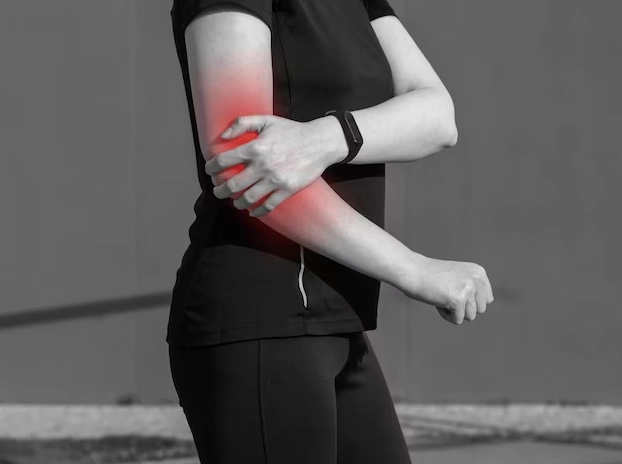
Dislocated elbow treatment in Hyderabad
Elbow Dislocation – Symptoms, Diagnosis & Treatment
Here is the active-voice version, with the passive constructions removed and the grammar cleaned up:
An elbow dislocation occurs when the radius and ulna in the forearm move out of their normal alignment with the humerus in the upper arm. These three bones form the elbow joint, and a dislocation occurs when they lose their normal alignment.
An elbow dislocation can also cause serious injuries, including fractures, damage to the arteries that carry blood to the hand, nerve damage around the elbow, and problems with movement or sensation in the arm and hand.
Causes of Elbow Dislocation
Most elbow dislocations occur after a fall, usually when a person lands with the arm fully extended.
This removes passive constructions such as “are displaced,” “can be dislocated,” “that can occur,” and “are caused by.”
But any trauma, such as a car accident, can cause an elbow dislocation.
Elbow Dislocation Symptoms
- Swelling
- Severe elbow pain
- Weakness in the joint
- Deformed looking arm
- Bruising
- Loss of ability to move the elbow
- Inability to bend the arm
Personal Care at Home
Elbow dislocation is a serious injury that requires medical help. However, you can take some self-care measures at home to ease swelling and pain. For example, you can apply ice to the elbow to help reduce swelling. Afterward, arrange an appointment with an orthopedic doctor for proper evaluation and treatment.
This Simple Test at Home Can Help You Check Whether the Artery and Nerves Are Intact
First, you can check the artery by feeling the base of the wrist below the thumb to locate the pulse. Similarly, press your fingertips gently; they should turn white and return to a pink color within a few seconds. If either test produces an abnormal result, seek medical attention immediately.
Nerve Function Test
In addition, three nerves pass through the elbow, and each nerve plays a role in strength and sensation. First, test your strength by flexing your wrist as if saying “stop” (radial nerve function). Next, extend your fingers to check ulnar nerve function. Finally, try to touch your thumb with your little finger to check median nerve function. If you experience any difficulty during these tests, contact your orthopedic doctor immediately.
Examination and tests
When you visit an orthopedic doctor, the doctor will examine you and perform a physical examination. First, the doctor will check your nerves and arteries by checking your pulse, testing sensation, moving your fingers and wrist, and confirming normal blood flow to your hand.
Next, the doctor will order an X-ray. In some cases, a fracture can look like a dislocation, and a fracture can also occur along with a dislocation.
If your doctor suspects an arterial injury, the doctor may order additional tests, such as an angiogram (X-ray of the arteries).
In addition, the doctor will test sensation by touching different areas of your hand and arm.
Dislocated Elbow Treatment in Hyderabad
During treatment, the doctor will reduce (realign) the elbow by gently retracting the wrist and guiding the joint back into place. Because this procedure can cause significant pain, the doctor may give you strong pain medication before starting the procedure.
When the elbow is back in place, the doctor will take an x-ray and put a splint on it to keep the elbow bent. The splint will form an “L” shape at the back of the elbow. It will be plaster or fiberglass. Its purpose is to prevent the arm from moving through the elbow. Your arms will usually be in a sling to help you hold up your splint.
Prevention
Take precautions and do not walk on slippery stairs and surfaces. Do not fall on outstretched arms. Avoid situations that make falls more common (such as walking at night or on slippery floors). Avoid overuse injuries by restricting overtraining in sports.
When to see a doctor? If you can’t move your elbow, have severe pain, can’t feel your hand, or feel uncomfortable, you should go to your doctor’s office or hospital emergency room right away. For the best dislocated elbow treatment in Hyderabad, meet Dr. Praharsha Mulpur.
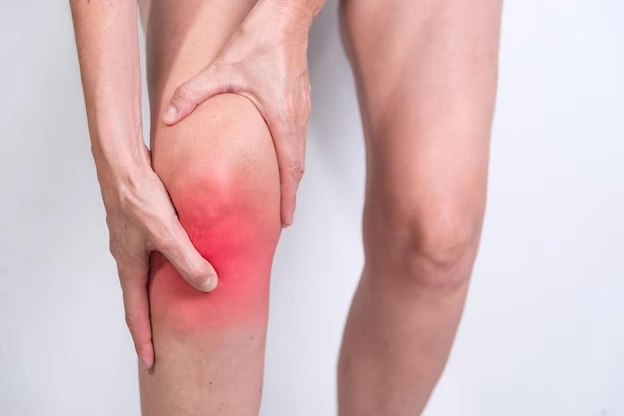
Tendinitis Causes Symptoms Treatment
Tendinitis Causes, Symptoms & Treatment
Tendinitis or tendonitis is a painful condition of the tendons (the connective tissue between your bones and muscles). Tendons are thick cords of connective tissues that join your muscles to bones. Tendinitis occurs in joints such as knee, hip, elbow, shoulder and at the base of the thumb (Achilles tendon). The term tendinitis or tendonitis is used interchangeably. In simple terms, tendinitis is the inflammation of tendons. The most common reason for this type of problem is repetitive activities and motions of the joint. In severe cases of tendinitis, moving the joint becomes difficult with acute pain and tenderness.
Tennis elbow, Golfer’s elbow, Pitcher’s shoulder, swimmer’s shoulder are the common examples of tendinitis.
Tendinitis causes, symptoms, treatment
Involvement in repetitive action or activity over a period of time may lead to tendinitis. For instance, making the same movement of your hands, legs, elbow joints over and over again can lead to tendinitis. The risk augments if you don’t perform your activity correctly.
Athletes are at higher risk – especially those who play basketball, bowling, tennis or golf.
People who are in jobs that require repetitive tasks or motions, overhead lifting, excessive physical work or exertion are at risk.
The risk of tendinitis increases with age, injury, rheumatoid arthritis and diabetes and also by the use of certain medicines.
Older people lead sedentary life with minimum activity. Aging and inactivity can lead to lower flexibility of tendons making them prone to wear and tear and injuries.
Tendinitis Symptoms
- Pain in and around the affected joint
- Dull achy joint
- Joint looks tender to touch
- Pain augments if someone touches the affected area
- Pain increases with movement
- Swelling
- Tightness makes it difficult to move the joint
Diagnosis
Your orthopedic doctor performs a physical examination of the affected joint. The doctor will also ask you about the symptoms and your medical history. Your doctor will also check tenderness and a range of motions. You should tell the doctor about any past or recent injuries, involvement in sporting activities or physical exercises, use of prescription or over-the-counter medicines. Based on the requirement, you doctor may order, X-rays, Ultrasound or MRI scan.
When to see your orthopedic doctor?
You can take care of mild tendinitis by yourself adopting recommended measures and home-based remedies. However, when symptoms persist and trouble you making your routine activities difficult, then you should consult your orthopedic doctor.
Treatment
Taking rest, heat or ice application, use of OTCs and doing exercises and stretches that help build, strengthen muscles and improve mobility will help.
In severe cases your orthopedic doctor may recommend use of supports such as cane, braces or splints, physical therapy and injections (corticosteroids). In extremely painful and severe cases, your doctor recommends surgery to remove inflammatory tissue.
Why you should treat Tendinitis in time?
Tendinitis complications: Symptoms can become progressively worse if they last long interfering with daily activities. The risk of tendon rupturing is very high, if you don’t treat it properly. And, if the tendon ruptures, surgery becomes mandatory. Tendinosis is a degenerative condition of the tendon associated with severe pain and inflammation. Tendinitis can lead to this condition if the symptoms left undressed for several weeks or months.
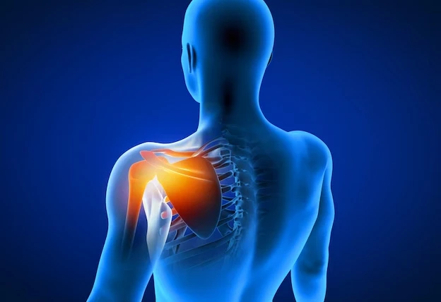
Frozen Shoulder – Symptoms Causes & Treatment
Frozen shoulder treatment in Hyderabad | Dr. Praharsha Mulpur
Adhesive capsulitis or Frozen shoulder – both these terms are used interchangeably. Frozen shoulder is a condition in which shoulder pain and stiffness are the prominent symptoms. The symptoms manifest rather slowly and then become worse over time. During this time, the affected shoulder becomes very difficult to move.
The symptoms altogether take a longer time to get better. People who develop frozen shoulders keep their shoulders stiff for a long time owing to which the condition becomes worse. Getting better from a worsened state usually takes a very long time.
Frozen Shoulder Develops in Three Stages
- Freezing Stage: During this first stage, the pain gradually becomes more and more intense. As the pain worsens, the shoulder loses its motion. A freezing stage may last for up to 45 days to 9 months.
- Frozen Stage: Pain eases somewhat during this period, but shoulder stiffness remains. This stage may last for up to 4 to 6 months. Performing daily activities may become a trivial task during this time.
- Thawing stage: Motion gradually starts to improve during this stage – and, it may come close to normal motion and strength – but, it may usually take around 8 months to two years.
Frozen shoulder Causes
There is no known cause of frozen shoulder as the causes are not fully understood. Even occupation or arm dominance doesn’t have any clear connection. However, there are some factors that can make a person at risk of developing a frozen shoulder:
Immobilization: When the shoulder is kept immobilized for a certain period of time after an injury, fracture, or surgery, a frozen shoulder may develop. Therefore, orthopedic doctors recommend their patients move their shoulders after surgery to prevent this condition.
Frozen shoulder symptoms
Shoulder pain is the most prominent symptom of a frozen shoulder. It is usually aching or dull pain that becomes intense when you move your arm. The outer shoulder feels painful and sometimes the upper arm as well. The other major symptom is a limited range of motion.
Diagnosis of Frozen Shoulder
Your doctor will discuss your medical history and symptoms and then examines your shoulder. Your orthopedic doctor will keenly observe whether you will feel pain when you move your shoulder. In this way, the doctor will check and compare both the active range of motion (when you move your shoulder on your own) and the passive range of motion (when someone else moves your shoulder). After making you move your shoulder in all possible directions your doctor will try to see whether your shoulder movement is restricted and whether you feel pain with motion.
Furthermore, to rule out other causes of your shoulder stiffness and pain, your doctor will order the following tests:
X-rays: To detect any structural issues and fractures in the bones and also problems such as arthritis, X-rays are helpful.
An Ultrasound and MRI: Soft tissue injuries can also cause pain and stiffness in the shoulder joints. To have clear and better images of soft tissues ultrasound and MRI tests are helpful. Though these tests don’t help in diagnosing a frozen shoulder, they help in identifying torn rotator cuff.
Frozen Shoulder Treatment in Hyderabad
Without treatment, the symptoms to improve may take a very long time – maybe up to 2 to 3 years. Therefore, the major goal of treatment is to control pain and restore strength and motion.
The treatment for a frozen shoulder involves medications, physical and occupational therapy with a range of activities and exercises, and the use of corticosteroid injections.
Primary treatment involves physical therapy with an aim to improve the flexibility of shoulder muscles.
Arthroscopic surgery is recommended in rare cases. This will help loosen the joint capsule so that it can move very easily.
The non-surgical treatment for frozen shoulder
Pain relieving medicines (NSAIDs) – Ibuprofen and aspirin help reduce pain, swelling, and inflammation.
Corticosteroid injections such as cortisone are directly injected into the shoulder joint. It is a powerful anti-inflammatory medicine.
Physical therapy – Some proven exercises help in restoring stiffness and motion. Physiotherapy is done under the supervision of a physical therapist through a home-based program.
Surgical Treatment
When typical conservative non-surgical treatment approaches such as medicines and physical therapy don’t offer any relief, then you should discuss with your orthopedic surgeon the scope for recovery while continuing with simple treatments and the benefits of considering shoulder surgery.
The goal of surgery is to stretch and release the stiffened joint capsule. Shoulder arthroscopy provides excellent outcomes.
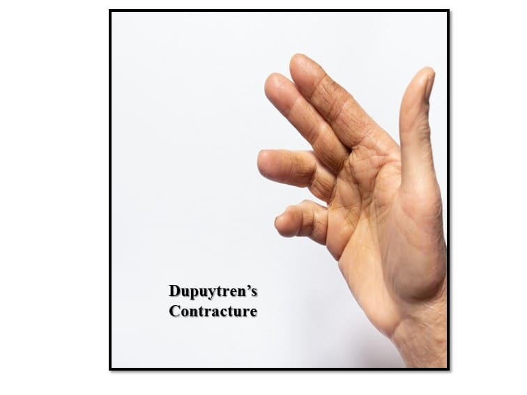
Dupuytren’s Contracture – Risk & Treatment
Dupuytren’s contracture is a condition in which fingers turn or bend towards the palm of the hand and do not straighten completely. In this painless condition, one or more fingers bend towards the palm.
Description of the Dupuytren’s Contracture
The fibrous layer of tissue that lies underneath the skin in the palm and fingers is known as fascia. It helps in anchoring and stabilizing the skin on the palm. Knots of tissues develop under the skin of the palm (fascia) near the fingers. In people with Dupuytren’s disease, the knots gradually become thick and develop into a thick cord – which eventually pulls fingers toward the palm of the hand.
What does it look like when you develop this condition?
Dupuytren contracture can occur in one or both hands. It develops gradually and becomes severe over years. Initially, the lump looks a little tender and firm but doesn’t cause any pain. The lump gradually becomes hard and extends up into the fingers. When it becomes tight, it pulls the fingers toward the palm – severely deforming the fingers.
What is the cause of Dupuytren contracture?
The cause of this condition is completely unknown. But genes ( family history) could play an important role.
What happens if you develop this condition?
The condition most commonly affects the ring finger and the little finger.
A person who develops this condition finds it difficult to perform day-to-day tasks such as knotting shoe laces, putting on gloves, placing hands in their pockets, or shaking hands.
Are there any risk factors for this condition?
The risk factors for Dupuytren contracture include the following:
Family history: The condition runs in families.
Age: It is most common after the age of 50 or above.
Diabetes: The risk increases if a person has diabetes.
Gender: males are prone to this condition and the symptoms may become progressively worse over time.
Occupation: People in certain occupations that involve extensive use of vibrating tools are at increased risk of developing this condition.
Orthopedic Doctor’s Examination
The doctor carefully examines the fingers and asks a few questions about the patient’s symptoms, medical history, and overall health. The doctor may examine and record the nodules and cord on the palm and test the feeling in the fingers. He also measures the range of motion of the fingers and thumb.
Treatment
Orthopedic doctors provide both surgical and nonsurgical treatments to help slow the progression of the condition, straighten the affected fingers and improve the motion of the fingers. Non-surgical treatment involves the use of steroid injections and splinting. If the contracture hinders the movement and function of the fingers, your doctor may recommend surgery. The goal of the surgery would be to straighten the affected fingers and improve the mobility of finger joints. Many patients show improvement in the movement of their fingers after surgery. If you think you have developed this condition, then meet Dr. Praharsha Mulpur.

Emerging Trends in Orthopedics
Robotic Orthopedic Surgery
Robotics is revolutionizing orthopedic surgery by providing amazing benefits to patients – such as minimal soft tissue damage, accurate positioning of the implant, a wide range of motion, and better stability of the joint.
The robotic arm is capable of controlling bone cuts by removing just enough bone with far better accuracy.
Accurate placement of implants using a robotic arm reduces future misalignment and repositioning. Surgical robots are more precise to do this job.
99% of patients feel complete satisfaction after robotic-assisted joint replacement surgery.
The robotic-assisted arm can perform many tasks including cutting and reshaping bones, positioning implants, guiding orthopedic surgeons in positioning sophisticated surgical instruments, and modeling joints in 3D.
Artificial Intelligence and Machine Learning in Orthopedics
Computers use a huge amount of data in real-time. They use this data to learn, adapt and improve (machine learning) and help deliver better orthopedic patient care.
AI helps accurately predict the surgical outcome of a procedure and, as a result, helps a robotic orthopedic surgeon plan appropriate post-operative care. In addition, machine learning models can accurately predict the outcomes of joint replacement surgeries.
Furthermore, AI-enabled tools can support remote patient monitoring (RPM) and remote therapeutic monitoring (RTM).
Benefits of Remote Therapeutic Monitoring (RTM)
Unlike remote patient monitoring, which measures physiological parameters, remote therapeutic monitoring measures non-physiological parameters such as pain levels and range of motion. As a result, patients can monitor their own health more effectively and engage more actively with their doctors, which may support better outcomes.
In addition, an AI-based remote therapeutic tool can use a mobile app and a smartphone camera to measure patient parameters such as range of motion and pain levels. Furthermore, doctors can set daily or weekly prompts that encourage patients to send personalized progress reports. Consequently, doctors can closely monitor patients’ progress and determine whether their recovery is progressing as expected.
Robotic Orthopedic Surgery Benefits in a nutshell
- Extremely high level of precision
- Smaller incisions and quicker healing time
- Patients can go back to work much sooner
- The risk of complications is minimal
- ROBOTIC JOINT REPLACEMENT
