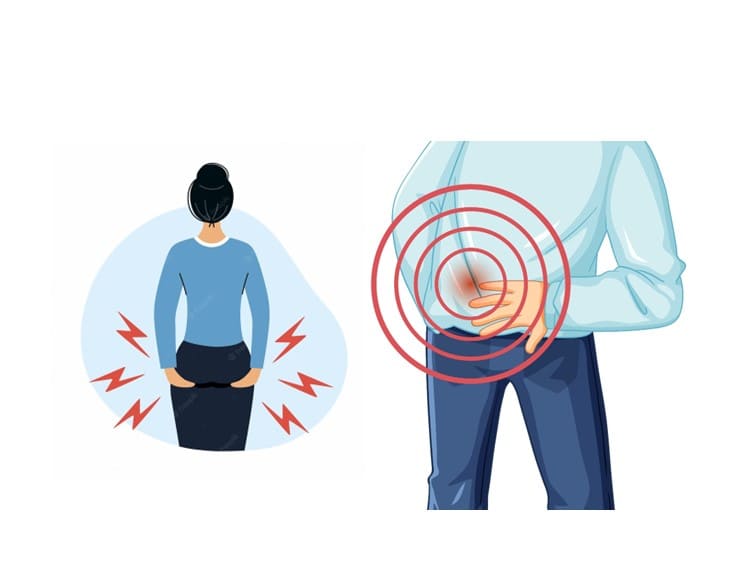
Can you explain what a hip labrum tear is?
Labral tear surgery Hyderabad
When someone tears the labrum of their hip, it means they have damaged the labrum, the cartilage in the hip socket. Your hip joint is a ball-and-socket joint that consists of a ball that rests on the femur (femoral head) and a socket that is part of the pelvis (acetabulum). The labrum helps keep the bones of the hip joint together and stable as you move. It also helps to hold the fluid together without friction. The degree of hip labrum tears varies. Sometimes there are small tears or abrasions on the edge of the hip labrum. This is usually caused by gradual wear and tear of the labrum. In other cases, part of the labrum of the hip joint may detach or be removed from the socket bone. This type of hip labrum injury is usually caused by trauma.
Hip labral tears can occur due to many reasons, although they are more common in athletes who participate in vigorous physical activity. Hip labral tears are commonly seen in women, as well as individuals with hip structure abnormalities such as hip dysplasia and other related medical conditions. Research indicates that up to 22% of athletes reporting groin pain are found to have hip labral tears.
What causes hip labrum tears?
Hip fractures can be caused by a number of factors, including
Fractures: Conditions that impair hip movement can cause fractures. In femoral vein entrapment (FAI), the femoral head does not fit into the glenoid socket. This failure can lead to chronic hip pain and fatigue. This is usually caused by chapped hips. FAI can affect people every day. If left untreated, it can cause osteoporosis in some patients.
Injury: A hip injury can cause a tear in the hip joint. This can happen in people who play repetitive, vigorous sports such as ice hockey, soccer, rugby, and golf. Degenerative health conditions: Osteoarthritis is the long (long) wear and tear of articular cartilage. As cartilage wears away gradually over time, it becomes more prone to tearing. Aging and obesity increase the risk of osteoarthritis. People with osteoarthritis often experience pain and stiffness in multiple joints, such as the hips and knees.
What are the symptoms of a labrum tear?
A tear in the acetabular labrum can result in a range of symptoms. While some individuals may not experience any discomfort, others may encounter severe pain around the groin area that may extend to the upper leg or buttocks. Pain may appear abruptly or slowly over time, and turning the leg can be particularly uncomfortable.
Symptoms of a hip labrum tear include: hip pain or stiffness; thigh or hip pain; hip pressed or locked when moving. Unstable feet when rotating the hip or when exercising or playing.
Acetabular labral tears may cause a sensation of the leg “catching” or “clicking” in the hip socket while in motion, and it may also feel like the leg is getting stuck or locked. Prolonged stress on the joint may lead to further degeneration and lasting damage.
It is also possible to develop an asymptomatic ischiatic labrum tear.
How is hip labral tear diagnosed?
To diagnose a hip labrum tear, your doctor will perform a physical exam. Your doctor may ask you to move your leg or walk around during the test. Your ability to move and any pain you experience while moving can help your doctor diagnose the condition. Imaging tests can also help doctors diagnose hip tears. Your doctor may order the following tests:
X-Ray: X-rays can alert your doctor to bone problems such as hip impingement or osteoporosis, which can cause labrum rupture and pain.
Magnetic resonance imaging (MRI): This test shows many things in Soft Tissue. An MRI can show the location and severity of the labral tear.
How is labral tear treated?
A hip tear will not heal on its own, but rest and other measures can help manage the symptoms of a minor tear.
Non-surgical treatments
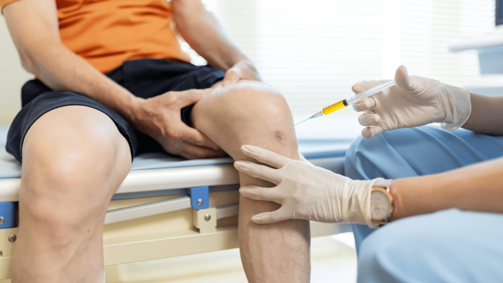
Over-the-counter pain relievers such as ibuprofen can reduce pain. Injections: Doctors may inject drugs such as steroids into the hip joint to relieve symptoms.
Physical therapy: Specific physical therapy that stretches and strengthens muscles can help reduce pain. Physical therapy usually requires a doctor’s prescription.
Hip Labral Tear Surgery in Hyderabad
If you have hip pain or have tried non-surgical options and still have pain, your doctor may recommend surgery. The most common surgery to repair hip pain is arthroscopic surgery.
Arthroscopic surgery
During this procedure, your orthopedic surgeon makes several small incisions to gain access to your hip joint. Using a special camera called an arthroscope, the surgeon can see the tear and repair it with surgical instruments.
When a hip labrum tear is caused by hip compression, the surgeon adjusts the bones in the hip joint to glide smoothly.
It is usually an outpatient procedure. This means you can leave the same day. After the hip labrum is repaired, people can resume normal activities, such as walking, almost immediately. The surgeon will refer you to a physical therapist who will begin exercises to restore joint strength.
If your job is in the office, you will be back to work in a week or two after a hip repair surgery. If your job puts a lot of strain on your hips, you can work with your physical therapist to determine a safe return date, or discuss with your doctor about changes in where you’ll be working so you can return to work. For the best labral tear surgery in Hyderabad, visit Ortho Pro Clinic.
Ankylosing Spondylitis Treatment in Hyderabad
Ankylosing Spondylitis (AS) is a chronic inflammatory disease that primarily affects the spine and sacroiliac joints. As a result, it can cause pain, stiffness, and, in some cases, severe disability. In addition, AS can affect peripheral joints such as the knees, ankles, and hips. Therefore, understanding the symptoms, causes, diagnosis, and treatment options can help people manage the condition more effectively.
What is Ankylosing Spondylitis?
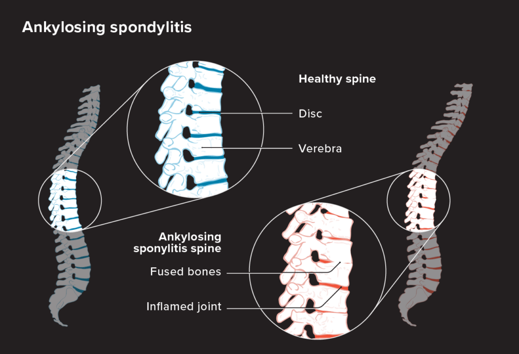
Ankylosing Spondylitis, often abbreviated as AS, belongs to a group of rheumatic diseases called spondyloarthritis. The term “ankylosing” refers to fused bones, while “spondylitis” refers to inflammation of the vertebrae or spinal bones.
AS primarily affects the spine and causes inflammation in the vertebrae. Over time, this inflammation can cause the spinal joints to fuse. Consequently, the spine can become stiff, less flexible, and more difficult to move. As a result, some people may find everyday activities increasingly challenging.
Symptoms and Diagnosis
Symptoms of Ankylosing Spondylitis
The symptoms of AS can vary from person to person. However, several symptoms commonly occur, including:
Chronic Pain: Persistent pain and stiffness can affect the lower back and buttocks, including the hip region. Typically, the pain becomes worse after rest and improves with movement.
Stiffness: Morning stiffness or stiffness after prolonged periods of inactivity can last for more than 30 minutes. Therefore, some people may find it difficult to move comfortably after waking up or sitting for a long time.
Fatigue: General fatigue and tiredness can occur because of the inflammatory process. In addition, ongoing symptoms can affect energy levels and daily activities.
Reduced Mobility: AS can make it difficult to bend or twist the spine. As a result, the condition can gradually limit the spine’s range of motion.
Other symptoms: People with AS may also experience tiredness, joint swelling, hip or thigh pain, neck pain or stiffness, and pain or stiffness around the ribcage. In addition, some people experience pain that becomes worse in the morning or after sitting for a long time. Foot, heel, or hand pain can also occur.
Causes and Risk Factors
The exact cause of Ankylosing Spondylitis remains unclear. However, researchers believe that a combination of genetic, environmental, and immunological factors contributes to its development.
In particular, the HLA-B27 gene has a strong association with AS. Nevertheless, having the HLA-B27 gene does not necessarily mean that a person will develop the condition.
Risk Factors
Several factors may increase the risk of developing AS. These include:
Genetics: A family history of AS or related conditions can increase the likelihood of developing the disease.
Age: AS typically begins during late adolescence or early adulthood. However, the condition can develop at other ages as well.
Gender: Men are more likely to develop AS than women, although the reasons for this difference remain unclear.
Diagnosis and Treatment
Diagnosing AS can sometimes prove challenging because its symptoms overlap with those of other conditions. Therefore, healthcare professionals usually consider several factors before confirming a diagnosis.
These factors include your medical history, physical examination, imaging studies such as X-rays and MRI scans, and laboratory tests. Together, these assessments help healthcare professionals evaluate the condition and determine an appropriate treatment approach.
Ankylosing Spondylitis Treatment in Hyderabad
Although Ankylosing Spondylitis has no cure, several treatments can help manage symptoms and improve quality of life. Depending on the individual’s condition, treatment may include medications, physical therapy, and lifestyle modifications.
Medications: Doctors may recommend nonsteroidal anti-inflammatory drugs (NSAIDs) to reduce pain and inflammation. In addition, they may prescribe disease-modifying antirheumatic drugs (DMARDs) or biologic therapies to target specific components of the immune system.
Physical Therapy: A structured exercise program can improve flexibility, strength, and posture. Furthermore, regular physical therapy can help maintain mobility and reduce the impact of stiffness.
Lifestyle Modifications: Maintaining a healthy lifestyle can also support overall management. For example, eating a balanced diet, quitting smoking, maintaining good posture, and practicing appropriate body mechanics can contribute to better physical function.
Surgical Interventions
In severe cases, conservative treatments may not provide enough relief. When this happens, an orthopedic surgeon may recommend surgery to address significant structural problems.
For example, spinal fusion surgery can help stabilize and realign the spine. In addition, during a spinal osteotomy, your orthopedic surgeon realigns the vertebrae to help you stand more upright.
During a laminectomy, your spinal surgeon removes part of a vertebra to relieve pressure on the spinal cord. Furthermore, if AS causes severe damage to the hip joint, your orthopedic doctor may recommend hip replacement surgery.
Living with Ankylosing Spondylitis
Living with Ankylosing Spondylitis can be challenging. However, understanding the condition and following an appropriate management plan can help you maintain mobility and manage symptoms.
Coping Strategies
Education: Learning about AS and its management options can help you take a more active role in your care. As a result, you may feel better prepared to discuss treatment decisions with your healthcare team.
Support Networks: Support groups and connections with other people living with AS can provide emotional support and practical advice. In addition, sharing experiences can help you manage some of the everyday challenges associated with the condition.
Self-Care: Prioritizing adequate rest, stress management, exercise, and relaxation techniques can support overall well-being. Furthermore, maintaining healthy daily habits can help you manage symptoms more effectively.
Bottom Line
Ankylosing Spondylitis is a complex inflammatory condition that can significantly affect mobility and quality of life. However, appropriate management can help people control symptoms and maintain function. Therefore, understanding the symptoms, causes, diagnosis, and treatment options can help you take proactive steps toward managing AS.
Ultimately, your treatment should match your symptoms, disease severity, and individual needs. If you experience persistent back, hip, or joint pain and stiffness, consult a qualified healthcare professional for an appropriate evaluation.
Injections, “Stem Cells” & Arthritis: What the Evidence Really Says
Regenerative injections for arthritis, including PRP, “stem cell” therapy, BMAC, and other biologic treatments, are increasingly marketed as ways to reduce pain, repair damaged cartilage, and delay joint replacement. But what does the evidence actually show? For patients with knee and hip arthritis, current research does not support claims that these injections can regrow cartilage in advanced, bone-on-bone osteoarthritis. Some treatments may provide limited or inconsistent symptom relief, while products marketed as “stem cell” injections may contain extremely small numbers of actual stem cells. If you are considering regenerative injections for arthritis, it is important to understand the evidence, potential benefits and risks, costs, and whether the treatment is appropriate for your stage of arthritis.
The Decision Many Patients Face
When knee or hip arthritis starts limiting daily life, it is completely natural to look for anything that might avoid or delay an operation. That instinct is reasonable — and exploring genuine alternatives first is exactly the right thing to do. The difficulty is that the internet, and some clinics, now offer a growing menu of injections promising to “regrow cartilage,” “heal the joint naturally,” or “avoid a knee replacement,” often for a large cash fee.
- You want to postpone or avoid surgery — and you deserve to know which alternatives are real.
- You’re told an injection can “regenerate” your joint — but is that actually true?
- It isn’t covered by insurance and can cost a great deal out of your own pocket.
- It’s genuinely hard to tell marketing from medicine when both sound confident.
Why the Marketing Can Mislead
- Much of the “evidence” promoted for these injections comes from small, industry- or provider-sponsored studies — not independent, high-quality trials.
- Online information often emphasises benefits while leaving out the risks, the costs, and the simpler alternatives that work.
- The label “stem cell” is routinely used for products that contain almost no stem cells at all.
Absolutely. I’d make the wording more active, direct, medically professional, and SEO-friendly, while keeping the evidence and terminology from your article unchanged.
What’s Actually Being Sold?
Several types of injections are marketed for knee and hip arthritis. Here’s what each treatment involves and what the evidence actually shows.
PRP
Platelet-Rich Plasma
What it is: A doctor draws a small amount of your blood and spins it in a machine to concentrate the platelets. The doctor then injects the concentrated plasma into the joint.
The evidence: In advanced knee arthritis, good-quality trials found no meaningful benefit compared with a saline injection, steroid injection, or gel injection.
BMAC
“Stem Cell” Injection
What it is: A doctor draws bone marrow from your pelvis, processes it to concentrate the cells, and injects the preparation into the joint. Clinics often market BMAC as a “stem cell” treatment.
The catch: Commercial BMAC preparations contain less than 0.01% actual stem cells. Therefore, calling BMAC a true “stem cell” treatment can be misleading.
“Stem Cells” / MSCs
From Fat or Lab-Grown Cells
What it is: Clinics obtain these cells from fat or bone marrow. Some providers also grow the cells in a laboratory before injecting them and market the treatment as a regenerative therapy.
The evidence: Studies show, at best, a mild benefit over placebo. Moreover, these treatments have not performed better than a simple steroid injection for advanced knee arthritis.
Viscosupplementation
Hyaluronic Acid “Gel”
What it is: A doctor injects hyaluronic acid, a lubricating gel, into the joint to help cushion it. Although it is not a biologic treatment, clinics often market it alongside PRP and other regenerative injections.
The evidence: Patients with advanced arthritis may experience only a small and inconsistent benefit. Consequently, several clinical guidelines no longer routinely recommend hyaluronic acid injections.
Stem Cell Therapy for Arthritis: What Does the Evidence Show?
<0.01%
Commercial BMAC preparations contain less than 0.01% mesenchymal stem cells. That represents an extremely small concentration and does not provide evidence that the injection can regenerate lost cartilage.
In simple terms, fewer than 1 in every 10,000 cells are mesenchymal stem cells. Therefore, patients should carefully question claims that BMAC injections can “regrow” damaged cartilage.
A Few Numbers Worth Knowing
Not All Cartilage Damage Is the Same
This is the single most useful idea to take away: whether anything can “repair” cartilage depends entirely on what kind of damage you have.
Focal Cartilage Defect
A localised “pothole” · often younger, after injury
Arthroscopy: the crater is the defect; surrounding cartilage is intact.
- The cartilage around it is still healthy.
- Genuine repair options exist — e.g. OATS (transplanting a small plug of your own cartilage-and-bone) or MACI (growing your own cartilage cells and re-implanting them).
- These are established, evidence-based procedures — for the right patient.
Advanced (Age-Related) Osteoarthritis
The arthritis affects the entire joint, often resulting in bone-on-bone contact.
X-ray: The joint space becomes narrow, and bone spurs (osteophytes) develop.
-
Advanced osteoarthritis destroys cartilage throughout the joint, leaving no localised area for doctors to repair or patch.
-
No injection or “stem cell” product can currently regrow an entire compartment of lost cartilage.
-
Instead, doctors focus on managing symptoms and maintaining function. When arthritis significantly affects quality of life, joint replacement can provide a reliable treatment option for the right patient.
What Genuinely Helps Arthritis
- Staying active, with targeted physiotherapy and muscle strengthening.
- Weight management — every kilogram lost meaningfully reduces load on the joint.
- Simple pain relief, and for occasional flares, a well-timed steroid injection.
-
When advanced arthritis starts affecting your quality of life, modern hip knee replacement offers one of the most reliably successful treatments in medicine. However, an orthopaedic surgeon recommends joint replacement only when it suits the patient’s symptoms, health, and stage of arthritis — not simply because arthritis appears on an X-ray.
Choosing the Right Arthritis Treatment
Importantly, cartilage damage varies from patient to patient, so doctors choose treatment based on the type and stage of arthritis. For example, carefully selected patients with localised cartilage defects may benefit from specific cartilage-repair procedures. In contrast, advanced age-related osteoarthritis destroys cartilage across the joint, and current injections or “stem cell” products cannot regrow the lost cartilage. In addition, targeted physiotherapy, muscle strengthening, weight management, appropriate pain relief, and selected steroid injections can help patients manage arthritis symptoms and maintain mobility. Ultimately, when severe knee or hip arthritis limits daily activities and affects quality of life, an orthopaedic surgeon may recommend joint replacement after considering the patient’s symptoms, X-rays, activity level, and overall health.
For advanced, bone-on-bone hip and knee arthritis, biologic injections — including PRP, “stem cells,” and BMAC — cannot currently be recommended for routine treatment. They have not been shown to regrow or restore lost cartilage in advanced osteoarthritis, and evidence of meaningful improvement over other treatments remains limited. — AAHKS Position Statement on Biologics for Advanced Hip and Knee Arthritis
test

Shoulder Fracture: Causes, Symptoms, and Treatment
A shoulder fracture is a common injury. Typically, patients across all age groups experience significant pain.
The shoulder is a complex joint made up of three bones: the clavicle (collarbone), the scapula (shoulder blade), and the humerus (upper arm bone). Usually, a fracture occurring in any of these three bones will lead to severe pain, loss of mobility, or other complications. Understanding the causes, symptoms, and treatment options is crucial for effective management and recovery. Before delving into shoulder fracture treatment options, we should explore their causes, types, and symptoms.
Causes of Shoulder Fracture
Shoulder fractures can result from various incidents, including:
- Falls: The most common cause, especially in older adults, is falling onto an outstretched hand or directly onto the shoulder.
- Sports Injuries: Contact sports (like football, hockey, and rugby) and activities with a high risk of falls (such as skiing or skateboarding) can lead to fractures.
- Trauma: Car accidents, bike accidents, or any significant blow to the shoulder area can cause a fracture.
- Osteoporosis: This condition weakens bones, making them more susceptible to fractures, even from minor falls or stresses.
Shoulder fractures are really painful and they cause immobility to your regular tasks. It is always helpful, if you could consult a doctor on time. It is generally not the same for every patient. But, there are situations where we must take diagnosis. If you shoulder pain is not relieving in an expected time frame, you have to do some exercises that can fix the pain. Phyio is a final option but not a first choice. An ortho Doctor can diagnose it better as they are knowledgable to treat the right thing. We at Hyderabad, treated many such patients for shoulder fractures. Probably, any ortho doctor gives the best advise after checking the patients. However, Dr. Praharsha Mulpur is specially trained on Orthopedics and has experience in surgeries.
Symptoms of Shoulder Fracture
The symptoms of a shoulder fracture can vary depending on the severity and location of the break. Common symptoms include:
- Pain: Severe and immediate pain at the site of the fracture.
- Swelling and Bruising: Around the shoulder and possibly extending down the arm.
- Deformity: The shoulder may appear out of place or have an abnormal contour.
- Limited Mobility: Difficulty moving the shoulder or arm, often accompanied by a grinding sensation.
- Tenderness: The area around the fracture may be extremely sensitive to touch.
Types of Shoulder Fractures
- Clavicle Fracture: This is the most common type of shoulder fracture. It typically results from a fall onto an outstretched hand or a direct blow to the shoulder.
- Scapula Fracture: Less common due to the protection provided by the chest and muscles, scapula fractures usually result from high-energy trauma.
- Proximal Humerus Fracture: This occurs near the top of the upper arm bone, often from a fall on an outstretched hand, especially in older adults with osteoporosis.
Diagnosis
To diagnose a shoulder fracture, an orthopedic doctor will:
- Physical Examination: Assess the shoulder for signs of deformity, swelling, and tenderness.
- Imaging Tests: X-rays are the most common imaging test to confirm the fracture. CT scans or MRIs may be used for more detailed images, especially if surgery is considered.
Shoulder Fracture Treatment Options
The treatment for a shoulder fracture depends on the type and severity of the fracture, as well as the patient’s overall health and activity level.
- Non-Surgical Treatment:
- Immobilization: Using a sling or shoulder immobilizer keeps the shoulder in place and allows the bones to heal.
- Pain Management: Over-the-counter pain relievers like acetaminophen or NSAIDs (e.g., ibuprofen) can help manage pain.
- Physical Therapy: Physical therapy is essential to restore movement and strength once the initial pain and swelling subside.
- Surgical Treatment:
- Open Reduction and Internal Fixation (ORIF): Metal plates, screws, or pins are used to stabilize the bone fragments.
- Shoulder Replacement: In severe cases where the bone is shattered or the joint is damaged, a shoulder replacement may be necessary.
Recovery and Rehabilitation
Recovery from a shoulder fracture can take several weeks to months, depending on the severity of the fracture and the treatment method used. Key aspects of the recovery process include:
- Rest: Adequate rest and avoiding activities that stress the shoulder are crucial.
- Physical Therapy: Gradual rehabilitation exercises to improve flexibility, strength, and range of motion.
- Follow-Up Care: Regular check-ups with the healthcare provider to monitor healing progress and adjust treatment as needed.
Prevention
While not all shoulder fractures can be prevented, some measures can reduce the risk:
- Fall Prevention: Use handrails, avoid slippery surfaces, and ensure proper lighting to prevent falls, especially for the elderly.
- Protective Gear: Use appropriate protective equipment when engaging in sports or activities with a high risk of shoulder injury.
- Bone Health: Maintain a diet rich in calcium and vitamin D, and engage in weight-bearing exercises to strengthen bones.
Prevention is a myth in terms for a fracture. Because, you don’t know whether you fell into a fracture by yourself. However, there are some security measures which you can consider while playing some sports. You can also take some advanced protection layers for your shoulders useful for your driving.
Conclusion
A shoulder fracture is a painful and often debilitating injury that requires prompt and effective treatment to ensure proper healing and recovery. Understanding the causes, recognizing the symptoms, and seeking appropriate medical care are essential steps in shoulder fracture treatment. With the right treatment and rehabilitation, most people can regain full function of their shoulders and return to their normal activities.
