Can you explain what a hip labrum tear is?
Labral tear surgery Hyderabad
When someone tears the labrum of their hip, it means they have damaged the labrum, the cartilage in the hip socket. Your hip joint is a ball-and-socket joint that consists of a ball that rests on the femur (femoral head) and a socket that is part of the pelvis (acetabulum). The labrum helps keep the bones of the hip joint together and stable as you move. It also helps to hold the fluid together without friction. The degree of hip labrum tears varies. Sometimes there are small tears or abrasions on the edge of the hip labrum. This is usually caused by gradual wear and tear of the labrum. In other cases, part of the labrum of the hip joint may detach or be removed from the socket bone. This type of hip labrum injury is usually caused by trauma.
Hip labral tears can occur due to many reasons, although they are more common in athletes who participate in vigorous physical activity. Hip labral tears are commonly seen in women, as well as individuals with hip structure abnormalities such as hip dysplasia and other related medical conditions. Research indicates that up to 22% of athletes reporting groin pain are found to have hip labral tears.
What causes hip labrum tears?
Hip fractures can be caused by a number of factors, including
Fractures: Conditions that impair hip movement can cause fractures. In femoral vein entrapment (FAI), the femoral head does not fit into the glenoid socket. This failure can lead to chronic hip pain and fatigue. This is usually caused by chapped hips. FAI can affect people every day. If left untreated, it can cause osteoporosis in some patients.
Injury: A hip injury can cause a tear in the hip joint. This can happen in people who play repetitive, vigorous sports such as ice hockey, soccer, rugby, and golf. Degenerative health conditions: Osteoarthritis is the long (long) wear and tear of articular cartilage. As cartilage wears away gradually over time, it becomes more prone to tearing. Aging and obesity increase the risk of osteoarthritis. People with osteoarthritis often experience pain and stiffness in multiple joints, such as the hips and knees.
What are the symptoms of a labrum tear?
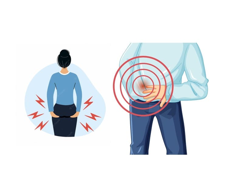
A tear in the acetabular labrum can result in a range of symptoms. While some individuals may not experience any discomfort, others may encounter severe pain around the groin area that may extend to the upper leg or buttocks. Pain may appear abruptly or slowly over time, and turning the leg can be particularly uncomfortable.
Symptoms of a hip labrum tear include: hip pain or stiffness; thigh or hip pain; hip pressed or locked when moving. Unstable feet when rotating the hip or when exercising or playing.
Acetabular labral tears may cause a sensation of the leg “catching” or “clicking” in the hip socket while in motion, and it may also feel like the leg is getting stuck or locked. Prolonged stress on the joint may lead to further degeneration and lasting damage.
It is also possible to develop an asymptomatic ischiatic labrum tear.
How is hip labral tear diagnosed?
To diagnose a hip labrum tear, your doctor will perform a physical exam. Your doctor may ask you to move your leg or walk around during the test. Your ability to move and any pain you experience while moving can help your doctor diagnose the condition. Imaging tests can also help doctors diagnose hip tears. Your doctor may order the following tests:
X-Ray: X-rays can alert your doctor to bone problems such as hip impingement or osteoporosis, which can cause labrum rupture and pain.
Magnetic resonance imaging (MRI): This test shows many things in Soft Tissue. An MRI can show the location and severity of the labral tear.
How is labral tear treated?
A hip tear will not heal on its own, but rest and other measures can help manage the symptoms of a minor tear.
Non-surgical treatments
Over-the-counter pain relievers such as ibuprofen can reduce pain. Injections: Doctors may inject drugs such as steroids into the hip joint to relieve symptoms.
Physical therapy: Specific physical therapy that stretches and strengthens muscles can help reduce pain. Physical therapy usually requires a doctor’s prescription.
Hip Labral Tear Surgery in Hyderabad
If you have hip pain or have tried non-surgical options and still have pain, your doctor may recommend surgery. The most common surgery to repair hip pain is arthroscopic surgery.
Arthroscopic surgery
During this procedure, your orthopedic surgeon makes several small incisions to gain access to your hip joint. Using a special camera called an arthroscope, the surgeon can see the tear and repair it with surgical instruments.
When a hip labrum tear is caused by hip compression, the surgeon adjusts the bones in the hip joint to glide smoothly.
It is usually an outpatient procedure. This means you can leave the same day. After the hip labrum is repaired, people can resume normal activities, such as walking, almost immediately. The surgeon will refer you to a physical therapist who will begin exercises to restore joint strength.
If your job is in the office, you will be back to work in a week or two after a hip repair surgery. If your job puts a lot of strain on your hips, you can work with your physical therapist to determine a safe return date, or discuss with your doctor about changes in where you’ll be working so you can return to work. For the best labral tear surgery in Hyderabad, visit Ortho Pro Clinic.

Ganglion cysts – Causes, Diagnosis & Treatment
Ganglion cyst treatment in Hyderabad – Dr. Praharsha Mulpur
manifest as masses that tend to emerge along the tendons or joints, primarily on the wrists or hands. It is also possible for them to develop in the ankles and feet. Ganglion cysts are usually spherical or elliptical in shape and are filled with a jellylike substance. They are not tumors
Ganglion cysts of a small size can resemble a pea. The size can alter. If a nearby nerve gets pressed, ganglion cysts can cause pain. Occasionally, they impact the mobility of the joints.
Features of Ganglion Cysts
Ganglion cysts typically exhibit these characteristics:
Location-The common location where ganglion cysts form is along the tendons or joints situated on the wrists or hands. Ankles and feet are next areas where ganglion cysts may form. Cysts can also develop in close proximity to other joints.
The shape of ganglion cyst is either circular or elliptical. Some cysts are so small to sense. Cysts tend to grow in size over time due to movements in the joint.
Pain-Typically, ganglion cysts do not cause any pain. In case a cyst applies pressure on a nerve or any other structures, it may result in muscle weakness, numbness, tingling, or pain.
When to See a doctor?
If you detect a lump or experience discomfort in your wrist, hand, ankle, or foot, it is advisable to consult an orthopedic doctor. The doctor will make a diagnosis to help assist whether management is necessary.
Causes
The reason behind a ganglion cyst remains unknown. A small water balloon-like structure on a stem originates from a tendon’s lining or joint to form the cyst. The cyst contains a viscous liquid that is similar to the one found in the joints or the areas surrounding tendons.
What are the risk factors for Ganglion Cysts?
Factors that can raise the likelihood of developing a ganglion cyst include:
Age and gender– Although anyone is susceptible to developing ganglion cysts, women aged 20 to 40 years may most often have them.
Osteoarthritis- Individuals with degenerative arthritis in the finger joints located closest to the nail beds have an increased likelihood of developing ganglion cysts in close proximity to those joints.
Tendon or joint injury– An injury to a tendon or joint increases the likelihood of developing a ganglion cyst.
Diagnosis
Your orthopedic doctor, during a physical examination, may apply pressure on the cyst to determine if it causes pain. To determine whether the cyst is solid or filled with fluid, your doctor shines it by passing a light through it. To confirm the diagnosis and exclude other conditions like arthritis or a tumor, your doctor orders imaging tests such as X-rays, ultrasounds, or MRIs. Confirming the diagnosis is also possible by extracting fluid from the cyst with a needle. Ganglion cyst fluid is usually clear and dense.
Ganglion Cyst Treatment in Hyderabad
If you have a painless ganglion cyst, with no other symptoms, your doctor may choose an observational approach where they monitor it and do not treat it unless necessary. Often, the cysts grow and shrink. Some go away on their own.
A ganglion cyst can grow in size with activity. Wearing a Brace or Splint can help because, they keep the joint still. It might help for a time to shrink the cyst and relieve pain and pressure on nerves. However, long-term use can weaken surrounding nerves and muscles.
If the cyst is hindering the mobility of your joint treatment is necessary. For a ganglion cyst that causes problems, an orthopedic doctor drains the cyst with a needle. If the cyst is causing severe problems, then it is removed surgically.
For the best ganglion cyst treatment, meet Dr. Praharsha Mulpur
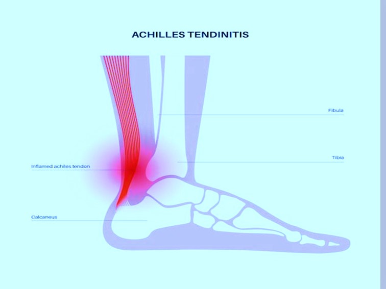
Achilles Tendinitis Types, Symptoms & Treatment
Achilles tendinitis is a common problem that occurs due to inflammation and irritation of the large tendon that runs down the back of the lower leg. This tendon, connects the calf muscles to the heel bone. It is responsible for facilitating several movements, including walking, running, climbing stairs, jumping, and standing on tiptoes. Despite its ability to endure significant stress from these activities, overuse can lead to tendinitis, which is a common condition associated with the Achilles tendon.
It is caused by overuse of the Achilles tendon, which is a band of tissue connecting the calf muscles to the heel bone in the lower leg. The injury is typically seen in runners who have suddenly increased the intensity or duration of their runs, as well as middle-aged individuals who participate in sports like basketball or tennis on weekends.
Description
Another term you might encounter is “tendinopathy,”. It describes a condition where the tendon experiences microscopic degeneration due to extensive damage over time. Tendinitis, tendinosis, and tendinopathy are interchangeable terms that describe the same issue.
Achilles Tendonitis Vs Achilles Rupture
Achilles tendinitis is not the same as Achilles rupture. The latter occurs when the tendon is either torn in half or separated from the heel bone. Achilles rupture is often caused by a sudden injury. The primary subject of discussion of this blog will be Achilles tendinitis rather than Achilles tendon rupture.
Symptoms of Achilles Tendinitis
- Achilles tendinitis causes
- Pain in the back of the heel (while wearing shoes)
- Persistent swelling in the affected area
- Formation of bone spur (insertional tendinitis)
- Thickening of the tendon
- Chronic pain after exercise
- Pain along the back of the heel or tendon that worsens with activity
- Pain and stiffness in the Achilles tendon
Achilles Tendinitis Types
There are two types –
- Nonintentional Achilles tendinitis
In this type the fibers located in the middle part of the Achilles tendon, that are just above where it attaches to the heel, are impacted. As time progresses, these fibers may start to deteriorate and form small ruptures, which can result in inflammation swelling and thickening of the tendon. This type of tendinitis is typically seen in younger individuals who are more active, particularly runners.
- Insertional Achilles Tendinitis
The condition known as Insertional Achilles tendinitis affects the lower part of the tendon where it connects to the heel bone or calcaneus. Over time, the damaged tendon fibers in both insertional and nonintentional Achilles tendinitis can harden (calcify) and form bone spurs on the heel, specifically in the case of insertional Achilles tendinitis. This condition can occur regardless of the activity level or time, although it is more prevalent among runners. Calf muscle tightness is a common cause of this condition as it increases the stress on the Achilles tendon insertion.
When to see a doctor?
If you experience any of the signs and symptoms associated with Achilles tendinitis – and also, experience severe and persistent pain along the Achilles tendon or the back of the heel, then you should see an orthopedic doctor immediately.
Orthopedic Doctor’s Examination
When you see a doctor and describe your symptoms, the doctor will examine your foot and ankle. During the physical examination, your doctor will see whether you feel pain while stretching your calf. The doctor will also look for these signs – swelling at the back of your heel; pain or bone spurs near the lower part pf the tendon; a reduced or restricted ability to point toes downward and pain in the middle of the tendon.
Treatment
Generally, when it comes to Achilles tendinitis, non-surgical treatments can effectively relieve pain, but it may take a few months for symptoms to disappear completely. Even with early treatment, the pain may persist for more than 3 months.
Anti-inflammatory pain medicines, corticosteroid injections, modifying activities, changing footwear, and doing physical therapy exercises are the primary methods used for non-surgical treatment.
Bottom Line
Most instances of Achilles tendinitis can be addressed through basic self-care methods with the oversight of a medical professional. It’s crucial to adhere to these approaches to avoid the condition from happening again. Nevertheless, for severe cases, Achilles tendinitis can lead to torn or ruptured tendons that may necessitate surgical treatment.
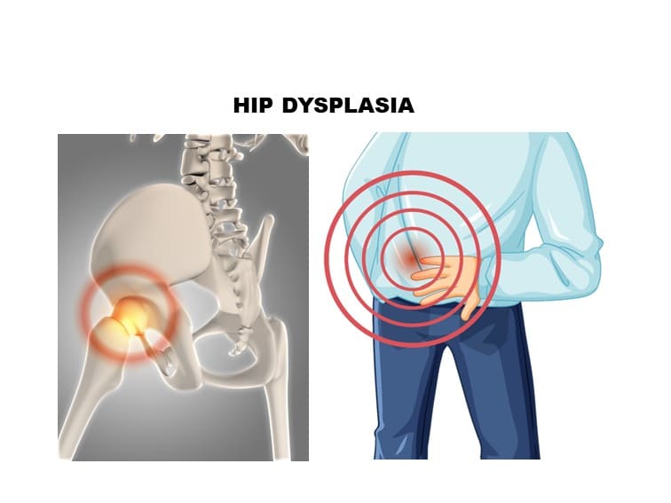
Hip Dysplasia Causes &Treatment
Hip Dysplasia Causes
The ball portion of the upper thigh bone fits into the hip socket and forms a stable ball and socket joint for smooth movement. If the socket doesn’t fully cover the ball portion, then the condition is known as hip dysplasia. It damages the cartilage lining the hip joint and the labrum (the soft cartilage) that covers the hip joint’s socket – hip labral tear.
This condition is mostly present in a child shortly after birth. However, if the condition is mild, then it doesn’t cause any symptoms until the child grows into a teenager or young adult.
Hip Dysplasia Symptoms
The signs and symptoms may vary according to the age group. During infancy, the mother can easily notice less flexibility in one hip than the other. The child limps as he or she begins to walk. One leg of the child seems to be longer than the other.
In teenagers and young adults, hip dysplasia can cause painful conditions such as hip labral tear or osteoarthritis. The pain in the hip and groin region increases with activity. Some persons may experience a sensation of instability in the hip joint.
Hip Dysplasia Causes
The problem mostly begins at birth. At this time of a person’s life, the hip joint is made of soft cartilage – which becomes hardened into bone. The ball and socket joint fit together perfectly as a mold. But when the ball fails to fit firmly into the socket – it remains shallow and does not fully form around the ball.
The hip joint moves out from its position during the last month before birth due to the space within the womb becoming crowded. The reduction in space in the womb may be due to the large size of the baby, first pregnancy, and breech presentation.
Hip Dysplasia Risk Factors
The risk increases if babies are born in the breech position. The condition is common in girls and also runs in families.
Complications
Hip dysplasia causes this condition: Hip dysplasia can lead to hip labral tear over time as it damages the labrum (the soft cartilage that rims the hip socket). The risk of osteoarthritis increases in people who have hip dysplasia.
Treatment
For children a variety of treatment options are available. The treatment depends on the extent of hip damage and the age of the child. For mild to moderate cases, orthopedic doctors treat the child with a soft brace. For babies older than 6 months, braces won’t work well. The orthopedic doctor repositions the bone in the proper position and then uses a full body cast to hold the hip joint in a proper position.
However, in severe cases, doctors correct the hip socket by cutting it free from the pelvis and then repositioning it so that it matches perfectly with the ball. The procedure is known as periacetabular osteotomy.
In older people, dysplasia can severely damage their hip joints leading to debilitating arthritis. For older people, hip replacement surgery remains an option.
Bottom Line
In older children, young adults, and older people, orthopedic doctors recommend surgery to move the bones into the proper position for proper movement of the joint.
Dislocated elbow treatment in Hyderabad
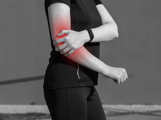
Elbow Dislocation – Symptoms, Diagnosis & Treatment
An elbow dislocation occurs when the bones in the forearm (radius and ulna) are displaced in relation to the bones in the upper arm (humerus). The elbow joint, that forms where these three bones meet, can be dislocated or separated.
The specific and serious injuries that can occur are fractures (broken arms), damage to the arteries in the arm (vessels that carry blood to the hand), damage to the nerves that run through the elbow area, and impaired movement and sensation in the arm and hands.
Causes of Elbow Dislocation
Most elbow dislocations are caused by a fall, usually with the arm is fully extended. But any trauma, such as a car accident, can cause an elbow dislocation.
Elbow Dislocation Symptoms
- Swelling
- Severe elbow pain
- Weakness in the joint
- Deformed looking arm
- Bruising
- Loss of ability to move the elbow
- Inability to bend the arm
Personal Care at Home
Elbow dislocation is a serious injury that requires medical help. You can take self-care at home to ease swelling and pain. To ensure this, put some ice on the elbow. Next, seek appointment of an orthopedic doctor.
This simple test at home can help you check whether the artery and nerves are intact
To check the artery, feel the base of the wrist below the thumb. This is to feel the pulse. Similarly, press your fingertips – they should turn white and return to pink color within a few second. If any of these tests are abnormal, seek medical attention immediately.
Nerve function test
Three nerves pass through the elbow. Each nerve contains sections that help increase strength and sensation. Test strength first by flexing your wrist as if to say “stop” (radial nerve function), then by extending your fingers (ulnar nerve function), then try to touch your thumb with your little finger (median nerve function). If you have any problems with these tests, meet your orthopedic doctor immediately. The
Examination and tests
When you meet an orthopedic doctor, he will see you and performs physical examination. The doctor will make sure that the nerves and arteries are not damaged by checking your pulse, making sure you feel normal, moving your fingers and wrist, and checking that the blood in your hand is flowing normally.
Next, the doctor must take an X-ray. Sometimes a fracture can look like a dislocation, and some fractures occur with dislocation as well.
If your doctor suspects damage to the arteries, other tests may be done, such as angiography (X-ray of the arteries).
Doctor will check sensitivity by touching the entire hand and arm.
Dislocated Elbow Treatment in Hyderabad
The doctor will reduce (realign) the elbow by retracting the wrist and lifting it back into place. It is very painful, so strong painkillers may be given before the procedure.
When the elbow is back in place, the doctor will take an x-ray and put a splint on it to keep the elbow bent. The splint will form an “L” shape at the back of the elbow. It will be plaster or fiberglass. Its purpose is to prevent the arm from moving through the elbow. Your arms will usually be in a sling to help you hold up your splint.
Prevention
Take precautions and do not walk on slippery stairs and surfaces. Do not fall on outstretched arms. Avoid situations that make falls more common (such as walking at night or on slippery floors). Avoid overuse injuries by restricting overtraining in sports.
When to see a doctor? If you can’t move your elbow, have severe pain, can’t feel your hand, or feel uncomfortable, you should go to your doctor’s office or hospital emergency room right away. For the best dislocated elbow treatment in Hyderabad, meet Dr. Praharsha Mulpur.