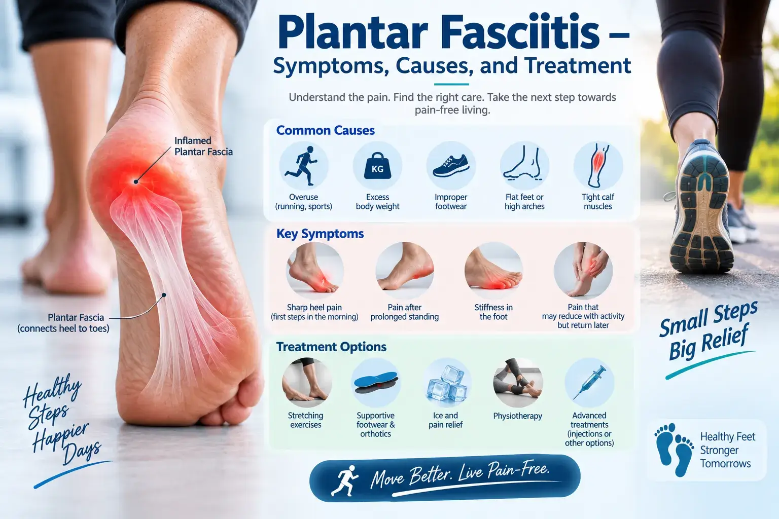
Plantar Fasciitis – Symptoms, Causes, and Treatment
Plantar fasciitis treatment in Hyderabad | Dr. Praharsha Mulpur
Many people experience heel pain. One of the common causes of heel pain is plantar fasciitis. It is due to the inflammation of a thick band of tissue called the plantar fascia. This tissue connects the heel bone to the toes. It runs across the bottom of each foot.
Plantar Fasciitis Causes
Plantar Fasciitis meaning: The thick band of connective tissue that runs across the bottom of each foot is known as fascia. It connects the heel bone to the base of the toes. It works like a shock absorber by supporting the arch of the foot and absorbing the load while walking.
Repeated stress and tension exert pressure on the tissue (fascia) and cause small tears. When these small tears expand and stretch repeatedly, they get damaged further. This may lead to irritation and inflammation of the fascia. However, in a majority of the cases of plantar fasciitis, the cause either remains unknown or is poorly understood.
Plantar Fasciitis Symptoms
Plantar Fasciitis Pain: People who develop this condition experience severe pain near the heel and in the sole (bottom) of the foot. They feel the worst pain early in the morning when they wake up and take the first few steps. They also feel pain after getting up from a sitting position. Standing for a long period of time can also trigger pain. The pain slows down and becomes less severe as a person gets up and begins to move.
Plantar Fasciitis Treatment in Hyderabad
Home care and lifestyle changes such as avoiding the activities that cause pain are helpful. Many people feel better and recover from planar fasciitis after a few months of conservative approaches such as stretching, applying ice packs, and modifying their activities. Pain medicines help in relieving pain and inflammation.
Plantar Fasciitis Exercises
Taking the help from a professional physical therapist can help in managing the condition better with a faster recovery. You can learn different exercises to strengthen the Achilles tendon and plantar fascia. Exercises also help strengthen your lower leg muscles. Your therapist will also teach you the use of taping to support the bottom of your foot.
Custom-fitted arch supports (orthotics) help in the even distribution of pressure on the feet. Your Orthopedic doctor may prescribe such off-the-shelf products. Similarly, your doctor may also recommend you wear a splint. It will help in holding the Achilles tendon and plantar fascia in a lengthened position and promote stretching while you sleep. To help reduce strain and pressure on your affected foot your orthopedic doctor may recommend using crutches, canes, or boots for a brief period of time.
Plantar Fasciitis Treatment in Hyderabad (Surgery)
Your orthopedic doctor may recommend the following therapies if the above measures fail to provide any relief: Steroid injections (the use would be limited) and PRP therapy; ultrasound tissue repair and extracorporeal shock wave therapy. Surgery may be recommended only in a few failed cases of conservative treatment and also when the pain becomes chronic.
Bottom line
Many people tend to ignore plantar fasciitis. Overweight people and runners are prone to this condition although why it occurs in some people is still not understood completely. Anyway, if you have this condition, you should never ignore it as it may lead to severe heel pain. You may not be able to perform your regular activities if you develop chronic heel pain. However, if you try to avoid pain, you may tend to walk differently by making changes to your foot position. When you try to change your walking style and foot position while walking, you are at risk of developing knee, back, and hip problems.
To know more about all the possible treatment modalities and the most effective Plantar Fasciitis Treatment in Hyderabad, consult Dr. Praharshamulpur.
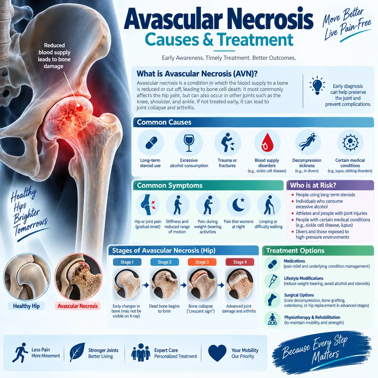
Avascular Necrosis Causes & Treatment
Avascular necrosis causes | Dr. Praharsha Mulpur Explains
Lack of blood supply to a bone results in the death of bone tissue. This condition is known as avascular necrosis or osteonecrosis. The condition can lead to minute breaks in the bone – which gradually cause the bone to collapse over a period of time. The process of bone damage may take several months to years. When a bone breaks or a joint dislocates, it can lead to blockage of blood flow to a section of the bone. Excessive and long-term use of certain medications, high-dose steroids, and alcohol can also cause avascular necrosis. Avascular necrosis can affect anyone, but it is most common in people between 30 to 50 years of age.
Avascular necrosis Causes
When the blood supply to a bone or joint is reduced, interrupted, or blocked – avascular necrosis occurs. The cause of a reduced blood supply can be:
Injury or trauma: bone fracture, bone injury or trauma, or a dislocated joint can damage nearby blood vessels. Radiation therapy can also weaken bones and damage blood vessels.
Medical conditions such as Gaucher’s disease and sickle cell anemia can reduce blood flow to bones.
Fatty deposits in blood vessels: lipids and fats accumulate and lead to the formation of plaque that blocks small blood vessels and reduces blood flow to bones.
Medical conditions associated with avascular necrosis include:
Sickle cell anemia, Systemic lupus erythematosus, HIV/AIDS, Gaucher’s disease, Pancreatitis, Certain types of cancer, such as leukemia and divers’ disease (Decompression sickness)
Symptoms
Avascular necrosis can affect the hip, thighs, buttocks, groin, shoulder, knee, hand, and foot. The condition develops gradually and can cause mild to severe pain. In cases involving the hip, patients typically feel pain around the buttock, groin, and thigh. Avascular necrosis of the hip and knee is common. It can affect one or both hips or knees.
Complications
Avascular necrosis can progressively become severe and worsen over time. As the condition progresses, the bones can become weak and eventually collapse. Furthermore, untreated avascular necrosis can lead to severe arthritis and significantly affect joint function.
Diagnosis
To determine the extent of the condition, your orthopedic doctor will perform a physical examination by pressing on your joints and checking for tenderness. In addition, the doctor will move your joints through different positions to determine whether you have restricted joint movement.
Furthermore, imaging tests can help your doctor identify changes in the affected bone.
X-rays: In particular, X-rays can help detect bone changes during the later stages of avascular necrosis.
In the early stages, X-rays usually don’t reveal bone changes or any issues in the bones.
CT and MRI Scans: These scans show early changes in the bones that are indicative of avascular necrosis. MRI and Ct scans produce detailed images.
A bone scan after injecting radioactive material into a vein helps in spotting the damaged or injured parts of the bones. On an imaging plate, these spots appear as bright spots.
Treatment
Early detection of the condition helps in giving effective treatment in the initial stages of bone damage. Doctors prescribe over-the-counter pain-relieving medicines and drugs to slow down the progress of avascular necrosis. If the cause of blockage is excess cholesterol and fat deposits then cholesterol-lowering drugs and blood thinners are prescribed. Taking rest as advised by the doctor and exercising under the supervision of a physiotherapist help maintain or improve movement in the joint.
Surgical treatment: Core decompression, bone reshaping (osteotomy), bone transplant (graft), and joint replacement are the surgical treatment options.
Bottom Line
You should consult an orthopedic doctor for any ongoing joint pain – especially when the pain is persistent. You must immediately seek emergency medical care for a possible dislocated joint or broken bone.
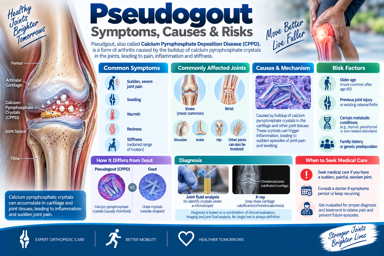
Pseudogout Symptoms, Causes & Risks
Pseudogout causes & Symptoms | Dr. Praharsha Mulpur
Pseudogout or calcium pyrophosphate deposition disease (CPPD) is a condition in which you have sudden and painful swelling in one or more joints. It can last for around a few days to weeks. The condition is commonly known as pseudogout because it looks similar to gout. The other common feature of both these conditions is the deposition of crystals within a joint though the type of crystals differs for each condition.
What are the symptoms of pseudogout?
The signs and symptoms associated with pseudogout include warm, swollen, and severely painful joints. The episodes may last for a few days to weeks. Pseudogout mostly affects the knees. It may also affect other joints, but ankles and wrists are affected less often.
What are the causes of pseudogout?
Pseudogout causes: The most prominent reason for this condition is due to deposition of calcium pyrophosphate dihydrate crystals in the affected joint of the body (most commonly in the knee joint). The condition is mostly associated with advancing age as older adults and people above age 65 to 70 years develop pseudogout. However, a majority of elderly people who have crystals in their joints never develop this condition. In addition, some people may have symptoms and others don’t have any symptoms at all.
What are the risk factors for pseudogout?
Age is a major risk factor. And also, the condition runs in families (genetic predisposition). This means people have a strong hereditary tendency to develop this condition. In addition, any serious accidental injuries, joint trauma, or post-surgical injury may also increase the risk of developing pseudogout.
Pseudogout causes: Certain medical conditions – such as underactive thyroid or an overactive parathyroid gland and mineral imbalances (excess iron or calcium in the blood or magnesium deficiency) can also increase the risk.
Bottom Line
The deposition of calcium crystals in the joints can damage the joints and cause stiffness, restricted mobility, and severe pain. The symptoms associated with pseudogout mimic the signs and symptoms of osteoarthritis or rheumatoid arthritis.
You should meet Dr. Praharsha Mulpur if you have sudden and severe pain, warmth, and swelling in your joints – especially in your knee joints. The symptoms may also be due to other joint conditions. Therefore, to rule out other joint conditions, a comprehensive physical and clinical examination and evaluation of the condition based on the symptoms is important.
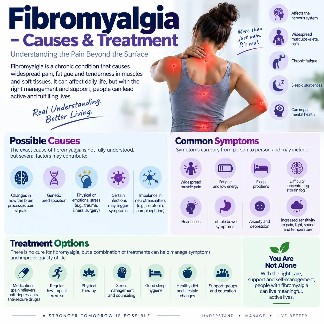
Fibromyalgia – Causes & Treatment
Fibromyalgia treatment in Hyderabad | Dr. Praharsha Mulpur
Fibromyalgia is a condition characterized by widespread musculoskeletal pain associated with fatigue, sleep, memory, and mood problems. Researchers believe that fibromyalgia increases painful sensations by affecting how the brain and spinal cord process painful and non-painful signals. Symptoms often begin after events such as Physical trauma, surgery, infection, or significant psychological stress. In other cases, symptoms gradually accumulate over time without a single triggering event occurring. Many people with fibromyalgia also experience tension headaches, temporomandibular joint disorders (TMJ), irritable bowel syndrome, anxiety, and depression. There is no cure for fibromyalgia, but various drugs can control symptoms. Exercise, relaxation, and measures to reduce stress can also help.
Fibromyalgia Symptoms
The main symptoms of fibromyalgia include:
Widespread pain
Fibromyalgia Symptoms
People with fibromyalgia often describe the pain as constant and dull, lasting for at least three months. Doctors consider the pain widespread when it affects both sides of the body and occurs above and below the hips.
Malaise: People with fibromyalgia may feel generally unwell or exhausted.
Sleep Problems: People with fibromyalgia may sleep for long hours but still wake up feeling tired. Pain can disturb their sleep, and many people also experience sleep disorders such as restless leg syndrome and sleep apnea.
Cognitive Difficulty: People often refer to this problem as “fibro fog.” It can affect concentration, attention, and the ability to focus on mental tasks.
Fibromyalgia often occurs alongside other conditions, including irritable bowel syndrome, chronic fatigue, migraines and other types of headaches, interstitial cystitis, and painful bladder syndrome.
Fibromyalgia Causes
Many researchers believe that repeated nerve stimulation in people with fibromyalgia can cause changes in the brain and spinal cord. These changes can increase the levels of certain chemicals in the brain that transmit pain signals. In addition, pain receptors in the brain may become more sensitive and develop a stronger response to pain signals.
Several factors may contribute to these changes, including genetics. Because fibromyalgia can run in families, certain genetic mutations may increase a person’s likelihood of developing the condition. Furthermore, some disorders may trigger or worsen fibromyalgia.
Physical or Emotional Events: Physical events, such as car accidents, can trigger fibromyalgia.
Persistent psychological stress can also trigger fibromyalgia.
Risk factors for fibromyalgia
your gender
Fibromyalgia is diagnosed more often in women than in men.
Family history- If you have a parent or sibling who also has fibromyalgia, you are more likely to develop fibromyalgia.
If you have osteoarthritis, rheumatoid arthritis, or lupus, you are more likely to develop fibromyalgia.
Complications
The pain, fatigue, and poor-quality sleep associated with fibromyalgia can affect your ability to function at home and at work. It can also lead to pain-related anxiety.
Diagnosis
In the past, doctors would check 18 specific points on a person’s body to see how much those points hurt when pressed hard. New guidelines from the American College of Rheumatology do not require a tender point test. Instead, the main factor required for a diagnosis of fibromyalgia is pain that has spread throughout the body for at least 3 months. To meet the criteria, there must be pain in at least 4 of these 5 areas: upper left region including shoulders, arms, or chin upper right region including shoulder, arm, or chin lower left region including hips, buttocks, or legs lower right region including hips, buttocks or legs. Axial regions including neck, back, chest, or abdomen test. Doctors may want to rule out other conditions with similar symptoms.
Blood tests include complete blood count, erythrocyte sedimentation rate, cyclic citrullinated peptide assay, and rheumatoid factor. Other tests include thyroid function tests, antinuclear antibody serology, and vitamin D test.
Fibromyalgia Treatment in Hyderabad
In general, fibromyalgia treatment includes both medication and self-care therapy. The focus is on minimizing symptoms and improving overall health. There is no cure for every symptom, but trying different treatment strategies can have cumulative effects. Medications can help relieve fibromyalgia pain and improve sleep. The common choices include over-the-counter pain relievers, muscle relaxants, and other prescription medicines. In addition, physical therapy, exercise, and other lifestyle and dietary modification can also help improve the condition.
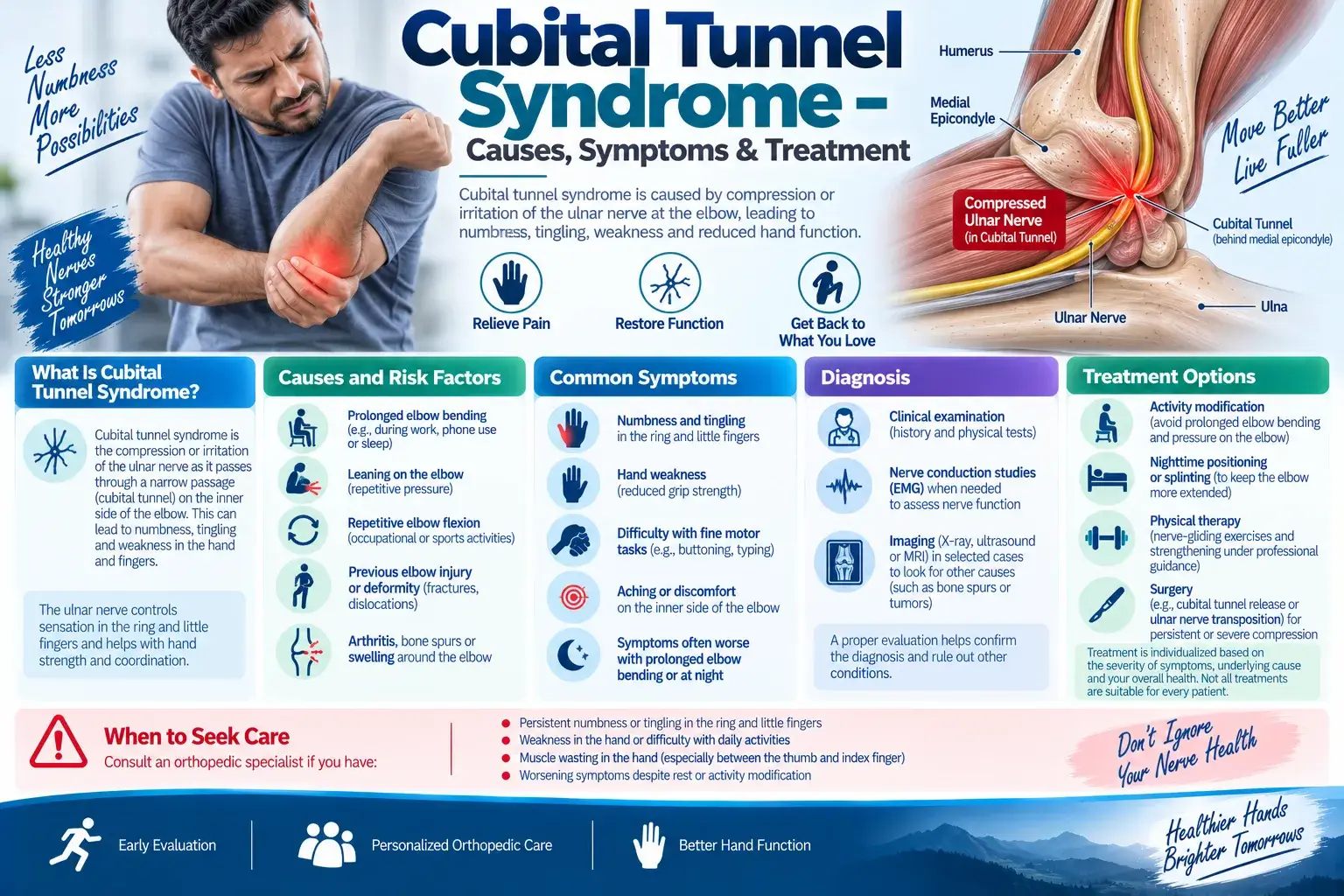
Cubital Tunnel Syndrome – Causes, Symptoms & Treatment
A nerve that travels from the neck to the arm and then to the fingers is known as the ulnar nerve. It is located near the ulna bone. It provides sensation to the ring and little fingers and to the forearm. The ulnar nerve extends the entire length of the elbow, forearm, and fingers. There is a small passage called the cubital tunnel through which muscles, ligaments, and ulnar nerve pass inside the elbow. This is the most common area where irritation may occur. The condition is known as cubital tunnel syndrome.
What causes cubital tunnel syndrome?
The cause of cubital tunnel syndrome is mostly idiopathic or unknown. However, many experts believe that excessive pressure, force, or tension created on this nerve due to repetitive movements may cause this condition.
The anatomical structure and positioning of the ulnar nerve with little soft tissue and a narrow space covering it make it susceptible to direct pressure. Therefore, any pressure or compression of this nerve causes numbness in the fingers.
Stretching – The nerve is vulnerable to stretching – especially during sleep.
Repetitive hand and wrist movements can also increase the risk of developing Carpal Tunnel Syndrome.
Cubital tunnel syndrome has different names – such as handlebar palsy, bicycler’s neuropathy, tardy ulnar palsy, ulnar nerve entrapment, or Guyon’s canal syndrome.
The following activities and any previous injury may put some people at a higher risk of developing cubital tunnel syndrome:
- Previous dislocation or fracture
- Swollen elbow joint
- Prolonged or repetitive movements
- Excessive flexing or bending of the elbow joint
- Arthritis or bone spurs of the elbow joint
What are the symptoms of cubital tunnels syndrome?
The ulnar nerve is a funny bone nerve. As this nerve is sparsely protected and close to the surface of the skin, it is more sensitive. If something hits in the inner elbow region, the sensation resembles an electric shock.
The signs and symptoms associated with cubital tunnels syndrome include:
- Stiffness in the fingers
- Problems manipulating things with hands or fingers
- Straightening and bending fingers are difficult
- Reduced grip or weakened grip strength
- Muscle loss at the base of the first finger and thumb
- Weakness in the arm, forearm, and fingers
- Pain in the arm, forearm, or fingers
How is cubital tunnel syndrome diagnosed?
Your orthopedic doctor may take note of the symptoms and perform a physical examination. The doctor will also take into account your complete medical history, and recommend the following tests:
X-ray: This test takes images of the bones and surrounding structures to see whether you have bone spurs or arthritis in your elbow.
Nerve conduction test: This test helps in determining the speed of signals that travel down a nerve. The test may be recommended to know whether any constriction or compression of the nerve is delaying the signals.
Electromyogram (EMG): This test helps in testing the forearm muscles controlled by the ulnar nerve. Both nerve and muscle functions are tested. When the muscles don’t work properly in their usual manner, it is an indication of a problem with the ulnar nerve.
Treatment
When you see your orthopedic doctor regarding the problem you may have, your doctor will instruct you to avoid certain actions that aggravate your symptoms, such as – leaning on the elbow, driving for a long time, resting your arms on the armrest of a chair for long; holding a book for a long time; typing extensively on a keyboard for long; holding a phone for a long time; and sleeping on the arm by bending the affected elbow.
In addition, your doctor will recommend the following home-based relieving methods:
- Using Pain relieving NSAIDs and anti-inflammatory medications and other OTC drugs
- Ensuring proper workstation ergonomics – the chair should not be kept lower than the tabletop
- Using an elbow pad that provides protection during the day
- Doing a thorough warm-up before exercise
- Resting the elbow and the arm whenever possible
- Regular stretching and strengthening exercises can help improve joint function and reduce the risk of repetitive strain injuries.
- Avoiding sports equipment or clothing that compresses the elbow
- Wearing an elbow pad during the day provides protection
- wearing an elbow splint at night to prevent the elbow from bending
- Wrapping the impacted arm loosely with paddings, such as cloth, towel, or pillow
Surgery
If you don’t get any relief from the nagging symptoms and the symptoms last longer than six weeks, your doctor may recommend surgery. Orthopedic doctors recommend surgery if your symptoms become extreme and chronic and if you experience weakness or muscle loss in your hand due to cubital tunnel syndrome.
