Foot Pain – Causes and Risk Factors
Foot Pain – Causes and Risk Factors
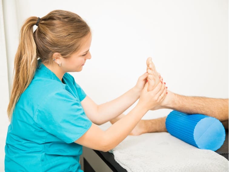
Foot pain causes
Your feet are important for you as they bear the entire weight when you move, walk, or wherever you go. They are prone to get strained or stressed if you overuse them. A majority of people don’t think much about foot pain until it hurt and causes limited mobility or movement. But, when your feet hurt, you want immediate relief.
Knowing the cause of the problem is important to get the right treatment. The location of the pain is also important to know the cause. Overuse, injury, or any other conditions that cause inflammation in tendons, ligaments, and bones can cause foot pain.
Your foot is the most complex part of your body. It has many muscles, joints bones, ligaments, and tendons. Foot pain is any discomfort or pain in one or more parts of your foot – such as soles, arches, heels, or toes.
Foot pain causes (Lifestyle Factors)
If you get foot pain, see whether you are wearing proper footwear. High-heeled sandals or shoes can cause foot pain as they put a great deal of pressure on the toes. This is, in fact, one of the prominent causes of foot pain in many people. And also check your activity levels including high-intensity exercises, intense aerobics, jogging, and weight-bearing exercises. These activities can also lead to foot pain if you get an injury during workouts.
What are the common medical causes of foot pain?
There are many medical problems associated with foot pain. Arthritis pain is the main cause of foot pain in many people. Arthritis can affect any of the 33 joints in the foot.
People with uncontrolled diabetes can develop several disorders and complications associated with the foot. They can become prone to:
- Foot sores or ulcers
- Nerve damage in the feet
- Hardened or clogged arteries in the feet and legs
Who are at risk of developing foot pain?
The following category of people are at risk for developing foot pain:
- Obese or overweight people
- Pregnant women
- People who have foot sprain, strain, fracture or foot injury, or tendinitis
What are the other potential causes of foot pain?
The other potential foot pain causes include:
Gout (commonly affects the big toe near the ball of the foot)
Fallen arches
Peripheral arterial disease (PAD)
Athlete’s foot
Hammer toes
Medications
Ingrown toenails
Warts
Bunions
Calluses
Corns
Haglund’s deformity (the back of the heel bone enlarges)
Morton’s neuroma (It is thickening around the nerve tissue between the toes near the ball of the foot)
When should you see your doctor?
If your foot pain triggers by certain activities, you must pay attention. If you know what triggers your foot pain, you will know better ways to manage it. However, you should not ignore your foot pain in the following circumstances:
- Your foot pain is due to a recent injury
- If the pain is sudden and severe
- You find it difficult to put weight on your foot.
- You have injured your leg owing to which you cannot put weight on your foot.
- If an open wound in your foot is causing pain
- The painful area has got discolored and has developed inflammation
- You have a fever in addition to foot pain.
- Your medical condition is interfering with blood flow due to which you are experiencing foot pain.
In the above cases, irrespective of whether you know your foot pain causes or not, you must see an expert orthopedic doctor to rule out any complex problem associated with your foot.

Medial Epicondylitis (Golfer’s or Baseball Elbow)
Golfer’s elbow causes | symptoms and treatment
You are well aware of tennis elbow. Golfer’s elbow or medial epicondylitis is quite opposite to the tennis elbow in terms of its effects. In this condition, you feel pain inside the elbow in the region of the bony bump due to cumulative damage and irritation in the tendons. These tendons attach to the muscles of the forearm. Whereas in tennis elbow, you feel pain outside of the elbow.
Is Golfer’s Elbow associated with Golfers Only?
Many people think that this condition is confined only to golfers, but it is not the case. Many active individuals and competitive athletes can develop this condition.
Forearm muscles and tendons help your wrist and fingers to move, twist and grip objects – such as a hammer, tennis racket, golf ball, or tennis ball. If you develop a golfer’s elbow, you may feel pain and difficulty gripping objects and playing. Affected individuals often experience pain or soreness at the inside of the elbow during or after activity.
What causes the golfer’s elbow?
Golfer’s elbow causes: Damage to the tendons and muscles that control wrist and finger movement causes golfer’s elbow (medial epicondylitis). People who are involved in forceful finger and wrist motions are subjected to repeated or excess stress. Repeated twisting and forceful gripping activities, poor conditioning, not doing warmup before any sporting activity; improper hitting, throwing, or lifting can lead to a golfer’s elbow.
What are the signs and symptoms of medial epicondylitis?
Golfer’s elbow symptoms: You may feel pain and tenderness in the funny bone region or in the bony bump. The pain is progressive and may radiate to the forearm. The pain can come on suddenly or gradually.
In the beginning, pain mainly occurs after activity, but may begin to interfere with activity. Once the pain starts to interfere, it will become difficult to pick up objects and grip door handles.
Another symptom is morning stiffness in the elbow or after a period of inactivity. You will feel stiffness when the elbow is fully straightened and slightly bend. Poor grip strength associated with pain
Golfer’s elbow causes and symptoms: Tingling sensation, numbness, and swelling are uncommon unless there is an injury. However, when the ulnar nerve that passes near the tendons gets irritated, you may feel pain, numbness, and tingling sensation in the fingers.
What is the treatment?
The goal of treatment is to identify and treat the cause (which is usually overuse or poor technique), reduce the pain and irritation, promote healing, and strengthen the muscles and tendons to prevent a recurrence.
When to see a doctor?
The first line of care is the use of over-the-counter pain relievers, the application of ice packs, and taking rest. Even after taking these measures if the pain and tenderness don’t ease, then seek a consultation with an orthopedic doctor who specializes in sports medicine. You must immediately seek care if:
- Elbow pain is severe and you can’t bend your elbow
- The elbow looks deformed
- Your elbow has swelling
- You have a fever with a swollen, hot, and inflamed elbow
- You suspect you’ve broken a bone

Hip Fractures Types, Treatment and Recovery
Hip Fracture Treatment in Hyderabad | Dr. Praharsha Mulpur
When the hip bone breaks hip fracture results. It mostly happens in the upper part of the hip bone (femur). Hip bone is the strongest bone, but can become weak in the elderly people due to osteoporosis. Therefore, even young people can become prone to hip fractures. In young people hip fractures can be due to severe fall and collision events – such as vehicle accidents.
The Hip Joint
Hip joint is a ball-and-socket joint. The upper part of the femur (thighbone) is the head (the ball). The part of the pelvis bone that is socket like is called acetabulum (rounded shape that fits around the femoral head).
Hip fracture types
Hip fractures due to injuries can occur in any of the following areas of the upper femur:
Femoral head: It is the ball of the femur that sits in acetabulum. The fractures of this type are extremely rare and may occur only when there is a high-impact or high-velocity even -such as road accident.
Femoral neck: The area below the femoral head is the femoral neck. This type of fractures are the common types of hip fractures.
Intertrochanteric area: The area is above the femur shaft and below the neck of the femur. It is called intertrochanteric because it is between two bony landmarks: the lesser trochanter and the greater trochanter. This type is also the most common type.
Subtrochanteric area: The upper part of the shaft of the femur below the greater and lesser trochanters.
Hip Fracture treatment in Hyderabad
Hip fracture treatment surgery Hip fractures require immediate surgical intervention as early as possible to lessen the risk of complications. Prior to surgery your orthopedic surgeon makes sure that you are medically fit. To ensure this, you will be seen by a cardiologist and pulmonologist. The process of getting ready for the surgery is called optimization. Preoperative testing should be done within 48 hours.
Hip fracture recovery
Physical and occupational therapists will work with the patient after surgery during the recovery period. They will show some exercises and tell the patient about the amount of weight they can put on the leg. They will also give instructions on how to manage daily activities – such as dressing and bathing.
Hairline hip fracture treatment
The first line home treatment or home remedy for a hairline fracture include RICE method – rest, application of ice, using compression and elevation. NSAIS’s are helpful to manage pain and swelling. If the pain and swelling doesn’t come down, you may need to see an orthopedic doctor. The treatment may depend on the extent, location and severity of bone injury. Your doctor may recommend using crutches to keep weight off the affected leg. Mild hairline fracture may heal on their own over a period of few weeks.
Hip fracture treatment without surgery
Isolated fracture of the greater trochanter: It occurs due to low-impact events such as household fall. This type of hip bone fracture is often painful, it usually heals without surgery. Patients with this type of fractures are treated with weight bearing aids such as walker and crutches.
Hip fracture in elderly
Hip fractures are not very common in young people. If they occur in young adults, they may be due to high-impact forces and events -such as fall from a height or motor-car or motor-bike accidents. In the elderly people bones become weak due to loss of mineral density related to age and osteoporosis and other health conditions.
Hip fracture X-ray
Most of the hip fractures can be diagnosed with X-rays.
MRI (Magnetic Resonance Imaging) Scans. For a more detailed visualization of fractures MRI provides fine images with complete details. It is helpful in detecting damage to soft tissues and bones. MRI scans are very sensitive as they can help detect an incomplete or small fracture that can not be seen on an X-ray.
CT scans – Computerized Tomography scans help provide detailed and clear images of the hip joint (cross-sectional images). An orthopedic doctor order a CT scan to get more details about the fracture.
Bottom Line
Painful hip fractures need immediate medical care and surgical intervention to fix the bones as early as possible. The primary goal of an orthopedic surgeon is to get the patient out of the bed so that complications such as blood clots, bed sores, infections and pneumonia can be prevented. When elderly people take prolonged bed rest, it may lead to disorientation and therefore their rehabilitation becomes very difficult. If you have any doubts pertaining to hip fracture treatment in Hyderabad, book an appointment with Dr. Praharsha Mulpur.
Soft Tissue Injuries – Part 2 by Dr. Praharsha Mulpur
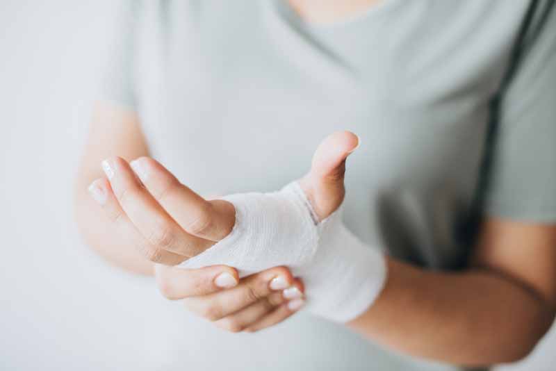
Soft Tissue injury treatment
Injuries to the soft tissues are known as soft tissues injuries. Muscles, tendons, ligaments are the soft tissues that are often prone to injuries due to excessive usage, strain, stress and repetitive movements of the joints such as shoulder, elbow, ankle and foot joints. In most of the cases, sudden injury or pain may result due to sudden abnormal or awkward movement of joints.
What is a stress fracture?
Excessive pressure, strain, force and usage may result in a stress fracture. It is a small crack or fracture in the bone. It mostly occurs in the weight bearing bones of the body – such as feet, legs, hip joint and knee joints. These are the weight bearing bones of the body. People who involve in excessive physical activity, sporting activity or any other type of physical work or laborious work are prone to stress fractures.
Stress Fracture (Soft Tissue Injury Treatment)
To address this type of fractures, one must stop the activity that causes the fracture, elevate the affected area, use pain medication and apply ice. The other treatment modalities that can be continued until you get relief may include minimizing pressure, force and stress on the affected joint, ceasing weight bearing activity, using shoe inserts, braces, casts and taking rest. In some cases, carelessness may lead to full blown cracks and fractures if small and simple crack progresses to a complete break. If this happens, surgery may be required.
What is bursitis?
There is a shock-absorber between the bones and the joints that connects two bones, muscles and tendons. It is a fluid-filled sac. Any injury, inflammation or infection of this cushion like sac may cause inflammation. It is known as Bursitis. It mostly happens due to overuse injury or direct injury to the joint. Bursitis mostly affects Hip, shoulder, knee (most commonly), elbow, foot and ankle.
What is tendonitis?
A band of soft tissues that connects muscles to bones are called tendons. Tendonitis is the most common type of soft tissue injury. It occurs when there is an injury or inflammation in the tendons. In other words, it is an inflammation of the tendons. The soft tissues – such as tendons when subjected to repetitive motion due to overuse – tendonitis may result.
Repetitive motions in sports such as badminton, shuttle, tennis, golf and others may trigger injury and inflammation. The tendonitis is thus named after the sports that trigger it – such as jumper’s knee, Golfer’s elbow, swimmer’s shoulder, and tennis elbow. Tendonitis commonly affects wrists, hand, elbow, knee, hip, shoulder, ankle and foot.
Soft Tissue Injury Treatment (Tendonitis)
If you have inflammation, pain and swelling, then you should first apply ice. You can do this immediately after the injury. Even cold compresses, elevating the affected joint, using anti-inflammatory medicines and taking rest can help in alleviating joint pain and inflammation. Orthopedic doctors recommend you to see a physiotherapist for stretching and strengthening exercises to prevent further injury to the joint. In some cases, if the pain becomes unbearable, doctors may give steroid injections. If a tendon is completely torn, surgery may be recommended.
Soft Tissue Injury Treatment
The first line treatment for getting relief from inflammation and other symptoms is taking anti-inflammatory medicines to relieve pain and reduce inflammation. Orthopedic doctors prescribe pain-relieving medicines and recommend application of ice to reduce swelling. Surgery is very rarely recommended. Physicians also suggest taking rest, keeping the affected joint elevated and using compressions as the first treatment modality. Doctors also prescribe antibiotics if they get a clue of infection in the affected joint.
Bottom Line
Both non-athletes and athletes share many similarities in soft-tissue injuries. If you have severe pain, swelling and inflammation due to a soft tissue injury then meet Dr. Praharsha Mulpur for a prompt and effective treatment.
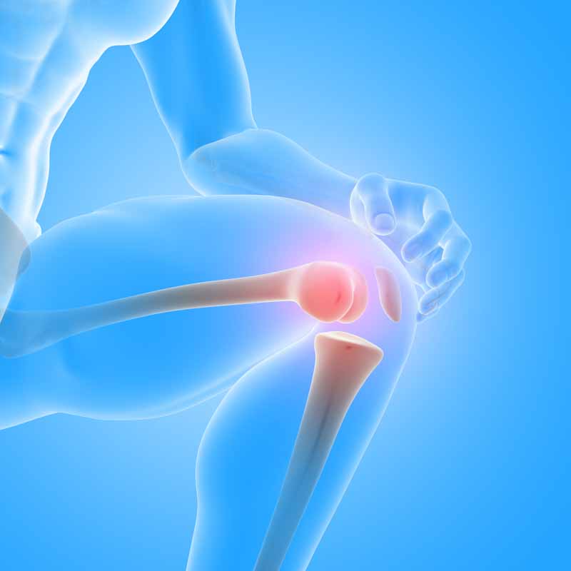
What To Expect After Knee Replacement | Dr Praharsha Explains
Care After Knee Replacement | Dr Praharsha
Care After Knee Replacement: Many people have these questions in their minds – What to expect after 3 months and after 5 months following knee replacement surgery. A majority of the patients recover after 3 months – and if it is a case of robotic knee replacement the recovery time is quite shorter. Nearly about 90% of the patients recovery almost 100% after 5 months following knee replacement. However, the recovery time may extent up to 6 months or longer in some cases. People with comorbid conditions may take longer time to recover. Older patients, smokers and individuals with other medical conditions may take longer time to heal. Recovery is faster in some people who perform knee joint and muscle strengthening exercises while they prepare for the surgery.
3 months after total knee replacement
Most people enjoy their routine activities such as walking comfortably, bicycling, playing games and even dancing at this stage after their knee replacement. You can also able to do a complete range of motions in your knee. Even with normal routine and recreational activities you don’t have any pain at all. However, the complete recovery still depends on your commitment and rehabilitation.
What not to do after knee replacement?
Don’t use bathtubs or slippery bathrooms because if you sit and bath it will be difficult for you to safely get up from there. Therefore, prefer bathroom with rough surfaces and do not try to reach to anything you need by bending or squatting even if you are in sitting position.
Forget about lifting weights and heavy stuff such as grocery bags or laundry or tool boxes, large pets and garbage bags. This will put excess stress on your knees.
What to wear after knee replacement surgery?
Wear comfortable footwear and shoes with non-skid soles. It is very important to wear footwear that provides additional friction on surfaces with anti-skid properties. Avoid wearing flip-flops as well. Avoid walking on wet and slippery surfaces. If you are new to a place then prefer walking slowly.
Equipment needed after knee replacement?
Your orthopedic surgeon may instruct you to use supportive devices that include canes, crutches and walkers. As long as your doctor recommend you to use assistive devices such as walkers use them. When you stand up on both legs use crutches and walker that help you to stand up. As far as driving is concerned ask your orthopedic surgeon about when can you start driving. Usually, you may have to wait for up to 5 to 6 weeks to resume driving for Care after knee replacement.
Sleep position after knee replacement?
Don’t bend your knees while sleeping or taking rest or when lying down. Taking pillow under your knees by bending them to support is also not recommended while taking rest or sleeping. It is better to keep your knee straight while you take rest or if you want to raise or elevate your leg.
How to take care of your new knee joint?
Long sitting is not advisable. Don’t sit in the same position for long – say for up to an hour or more – rather prefer getting up in between. Take breaks from sitting positions and long standing. Use sofas and chairs with armrests and a straight back. Don’t use rocking stools or rocking chairs or sofas and also chairs and stools without support and that are low.
If you are sitting in a chair or bed or sofa – don’t stand up abruptly from your sitting position. Take the support of armrest or bed support and then slide towards the edge and then get up. You can take the support of arms of the chair or walker or crutches to get up need Care after knee replacement.
When you are getting dressed
Don’t bend too much to dress and undress. Make yourself comfortable by sitting on the bed to get dressed without bending too much. Don’t dressed up while standing. While removing pants or clothes remove them first from the leg side that didn’t have surgery and from the surgery side last. While putting on pants and socks use the leg that has surgery done first.
Be Careful While Using Stairs
Care after knee replacement What precautions should you take as a part of care after knee replacement? Do not take long flight of stairs for up to two months after knee replacement. Use banister or side railing whenever you step up or down stairs. Do not try to move upstairs and downstairs as usual but take steps slowly until you are in complete control of your walking. This is important for your muscles to develop power and endurance. Use the leg that did not have surgery first while going upstairs. While getting down, first step with the leg that did have surgery.