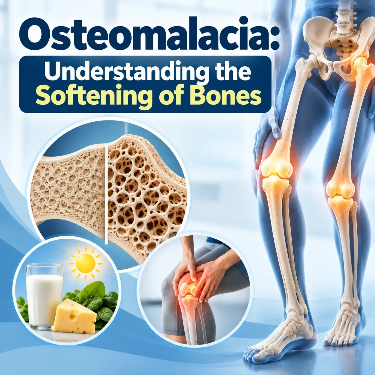
Osteomalacia: Understanding the Softening of Bones
Osteomalacia causes and treatment: is a metabolic bone disorder characterized by the softening of bones due to inadequate mineralization of the bone matrix. Unlike osteoporosis, which involves a decrease in bone density, osteomalacia primarily affects the mineralization process, leading to weakened and softer bones. This condition is often associated with vitamin D deficiency, calcium deficiency, or impaired absorption of these essential nutrients
Osteomalacia Causes and Treatment
1. Vitamin D Deficiency
One of the primary causes of osteomalacia is insufficient vitamin D. Vitamin D plays a crucial role in the absorption of calcium and phosphorus in the intestines. Without adequate vitamin D, the body struggles to maintain proper mineralization of bone, resulting in the softening of bone tissue.
2. Insufficient Calcium Intake
Calcium is a vital mineral for bone health. Inadequate calcium intake can contribute to osteomalacia as the body relies on this mineral for proper bone mineralization.
When your body lacks calcium, the bone matrix does not receive enough minerals, which can weaken the bones.
3. Malabsorption Disorders
Conditions such as celiac disease, inflammatory bowel disease, and certain surgeries, including weight loss surgery, can affect how your digestive system absorbs nutrients. As a result, your body may absorb less vitamin D and calcium, which can contribute to osteomalacia.
4. Renal Disorders
Kidney disorders can disrupt the body’s mineral balance. When kidney function declines, the kidneys may not convert enough vitamin D into its active form. Consequently, your body may absorb less calcium and phosphorus, two essential minerals for maintaining healthy bones.
Symptoms of Osteomalacia
Bone Pain: Osteomalacia can cause dull, aching bone pain, particularly in the hips, lower back, and legs.
Muscle Weakness: Osteomalacia can weaken the muscles because poor bone support can affect the muscles that help maintain skeletal stability.
Walking Difficulty: Weak bones can make walking difficult and increase the risk of fractures.
Diagnosis and Treatment
Blood Tests:</strong> Blood tests measuring levels of vitamin D, calcium, and phosphorus help diagnose osteomalacia. Low levels of vitamin D and calcium, along with elevated alkaline phosphatase, a marker of bone turnover, may indicate the condition.
Bone Biopsy: In some cases, doctors may perform a bone biopsy to directly assess the mineralization of bone tissue.
Imaging Studies: X-rays and other imaging studies can reveal characteristic bone density and structure changes associated with osteomalacia.
Treatment
Vitamin D Supplementation: The primary treatment for osteomalacia involves vitamin D supplementation. This may be administered orally or through intramuscular injections, depending on the severity of the deficiency.
Calcium Supplementation: Adequate calcium intake is essential for bone mineralization. Calcium supplements may be recommended to complement vitamin D therapy.
Management of Underlying Causes: If osteomalacia is secondary to malabsorption or renal disorders, addressing the underlying cause is crucial for effective treatment.
Prevention
Sun Exposure: Your skin synthesizes vitamin D when sunlight reaches it. Spending time outdoors can contribute to maintaining optimal vitamin D levels.
Dietary Intake:</strong> Consuming a diet rich in vitamin D and calcium, including fortified foods and dairy products, can help prevent deficiencies.
Regular Check-ups: Routine health check-ups, including monitoring of vitamin D and calcium levels, can aid in the early detection and prevention of osteomalacia.
Conclusion
Osteomalacia is a condition that highlights the intricate relationship between nutrition, bone health, and overall well-being. Recognizing its causes, symptoms, and proper management is essential for preventing complications and improving the quality of life for individuals affected by this metabolic bone disorder. Through a combination of adequate sunlight exposure, a balanced diet, and medical intervention, when necessary, it is possible to mitigate the impact of osteomalacia and promote stronger, healthier bones.
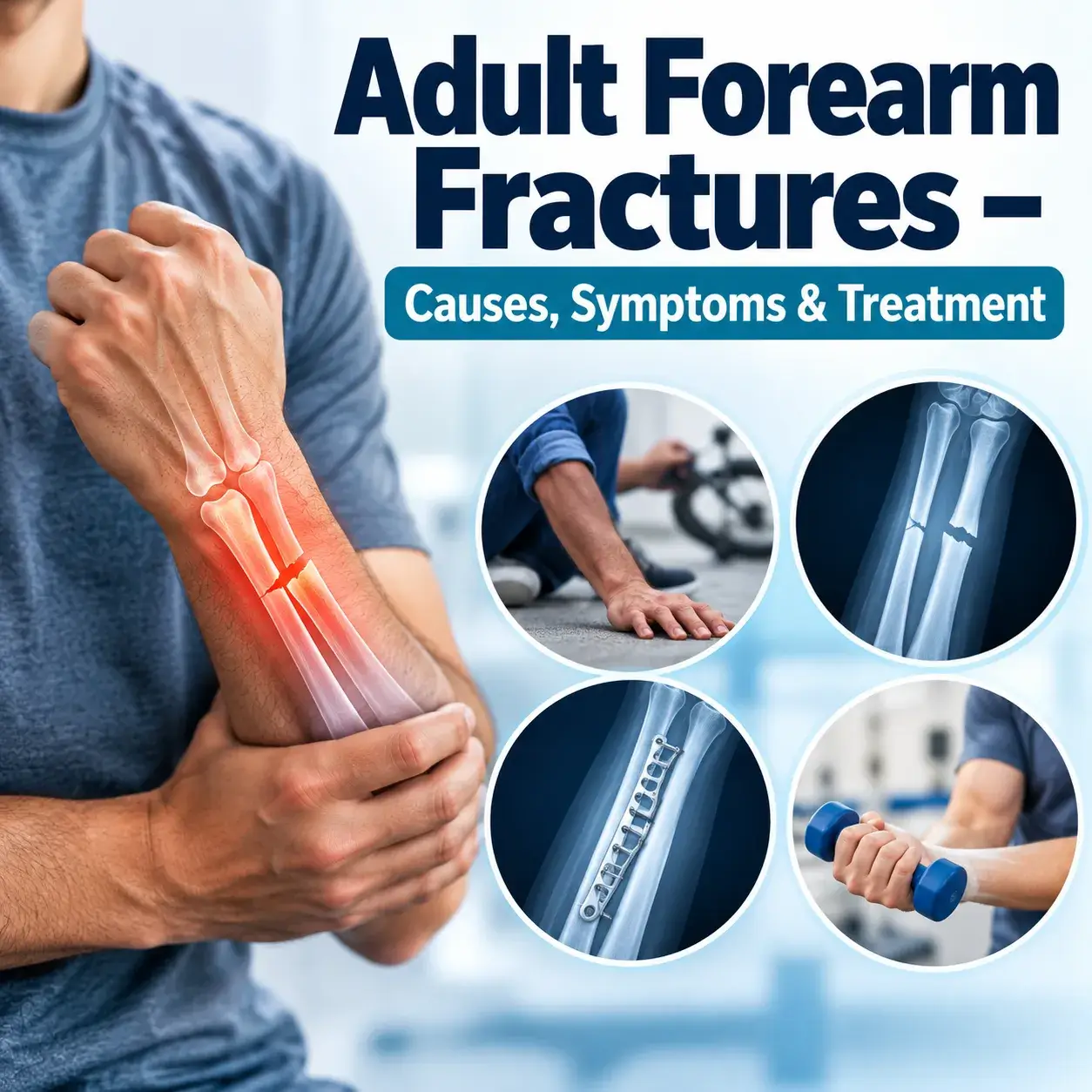
Adult Forearm Fractures – Causes, Symptoms & Treatment
Fractures of the adult forearm, though common, can have profound implications on daily life and functionality. The forearm, comprising the radius and ulna bones, is integral to a multitude of activities, and fractures in this region necessitate a comprehensive understanding of their types, causes, symptoms, and treatment modalities.
Forearm Fracture Types in Adults
Forearm fractures can manifest in various forms, with one or both bones affected. Isolated fractures of the radius or ulna are common, but a more complex scenario arises when both bones are involved. The position and extent of the fracture determine its classification, with common types including:
1. Radius Fracture: This often occurs due to a fall onto an outstretched hand or a direct blow. The fracture can be simple, with the bone breaking into two pieces, or comminuted, where the bone shatters into multiple fragments.
2. Ulna Fracture: Similar to radius fractures, ulna fractures result from trauma to the forearm. They can be transverse, oblique, or comminuted, depending on the nature and direction of the force applied.
3. Radius and Ulna Fracture: In cases where both bones are fractured, the alignment and stability of the entire forearm can be significantly compromised. These fractures often require careful evaluation and intervention.
Causes and Risk Factors
Forearm fractures commonly occur due to traumatic events, such as falls, sports injuries, or motor vehicle accidents. The vulnerability of the forearm bones to direct impact or torsional forces makes them susceptible to fractures during high-impact activities. Certain risk factors, including osteoporosis, can exacerbate the likelihood of fractures, as decreased bone density weakens the skeletal structure.
Symptoms and Diagnosis
The symptoms of a forearm fracture can be readily apparent, with pain, swelling, and deformity being key indicators. Limited range of motion, bruising, and tenderness at the fracture site are also common. A thorough clinical examination, coupled with imaging studies such as X-rays, is crucial for an accurate diagnosis. X-rays help in determining the type and extent of the fracture, aiding in the formulation of an appropriate treatment plan.
Treatment Options
The management of adult forearm fractures varies based on the type and severity of the injury.
In less severe cases, doctors may use conservative approaches such as casts or splints to immobilize the affected area. This approach allows the bones to heal naturally over time. However, more complex fractures may require surgical treatment.
Surgical options include open reduction and internal fixation (ORIF), in which the surgeon realigns the bones and secures them with screws, plates, or rods. For severe comminuted fractures or fractures that involve the joint, surgeons may use external fixation to stabilize the bones with pins connected to an external frame. The surgeon chooses the most appropriate technique based on the fracture pattern, the patient’s age, and overall health.
Rehabilitation and Recovery
Following treatment, rehabilitation plays a crucial role in restoring optimal function to the forearm. Physical therapy helps improve range of motion, strength, and flexibility. Gradual reintroduction of activities is essential to prevent stiffness and regain muscle strength. The duration of rehabilitation varies, with factors like the type of fracture and individual healing responses influencing the timeline.
Complications and Long-Term Considerations
While many forearm fractures heal successfully with appropriate treatment, complications can arise. These may include malunion, where the bones heal in a misaligned position, or nonunion, where the bones fail to heal. Nerve or blood vessel damage, though rare, can also occur. Long-term considerations involve monitoring for arthritis in the affected joint and addressing any residual functional limitations.
Bottom Line
In conclusion, adult forearm fractures present a multifaceted challenge that requires careful assessment and tailored intervention. Prompt diagnosis, appropriate treatment, and diligent rehabilitation contribute to optimal recovery and the restoration of functionality. Understanding the nuances of forearm fractures empowers individuals to actively participate in their recovery and ensures that the impact of these injuries on daily life is minimized.
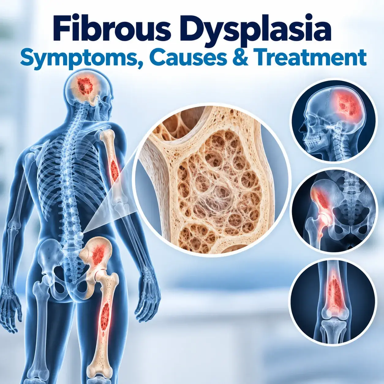
Fibrous Dysplasia Symptoms, Causes & Treatment
This is a type of bone condition wherein abnormal fibrous tissue develops in the normal bone replacing healthy bone tissue. This is a benign (noncancerous) bone condition. Over some time, fibrous tissue grows and spreads within the bone making it brittle and weak. A weak bone can become prone to fractures or deform.
In some people, the condition becomes severe affecting multiple bones and causing symptoms. Some people may experience a few or no symptoms at all.
When fibrous dysplasia becomes severe affecting multiple bones and causing severe symptoms, surgery becomes necessary to remove affected bones and prevent deformity or bone fractures.
Cause
Fibrous dysplasia is a rare genetic condition. It accounts for around 7% of all benign bone tumors. A gene mutation is responsible for making an abnormal type of fibrous bone.
Diagnosis
To diagnose this condition, your orthopedic doctor will take into account your medical history and overall health and ask you about your symptoms. The doctor will order X-rays and other tests to confirm the diagnosis of fibrous dysplasia.
X-rays – In a typical case of fibrous dysplasia. An X-ray may show:
Bone deformity – abnormal curvature or bowing
Expansion of affected bone
Abnormality in the affected bone
MRI or CT scan
An orthopedic doctor may order an MRI or a CT scan to further evaluate the lesion. An MRI scan clearly shows the extent of bone involved. It helps determine whether the lesion has turned cancerous.
A CT scan can help your doctor see the fracture better and determine the quality of the bone.
Bone scan: This scan helps in detecting additional lesions and abnormal bone structures in the body.
Treatment
Medications: Doctors prescribe medications and pain relievers to offer relief from pain and to decrease the frequency of fractures associated with weak bones.
Observation: If the condition doesn’t cause any symptoms, your orthopedic doctor may recommend monitoring and observation during regular follow-up consultations to check whether the condition is progressing.
Surgical treatment
Fibrous dysplasia is a progressive bone condition. It causes severe pain, weak bones, abnormal growths, and bone deformity. Your orthopedic doctor may recommend surgery to prevent bone fractures when you have progressive deformity of the bone; cracks in the bone; symptomatic lesions; displaced fractures; large lesions and lesions that have become cancerous.
Your orthopedic doctor scrapes a benign tumor out of the bone using a surgical procedure called Curettage to treat fibrous dysplasia.
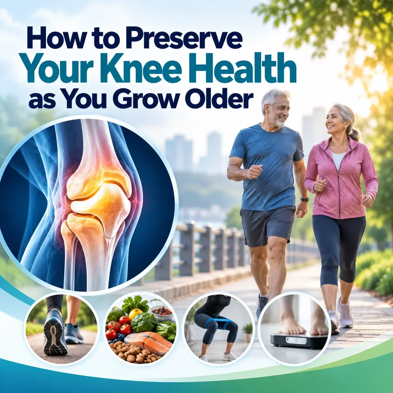
How to Preserve Your Knee Health as You Grow Older
As we age, it’s crucial to pay attention to our joint health, especially our knees. Knee problems can significantly impact our mobility and quality of life. In this article, we will discuss various strategies to preserve your knee health as you grow older, ensuring that you can stay active and pain-free.
Understanding the Importance of Knee Health
Why Are Knees Vulnerable
The knee joint is a complex structure, making it vulnerable to wear and tear.
Aging leads to the natural breakdown of cartilage, a crucial component of the knee joint.
Ensuring the well-being of your knees is crucial for your overall health.
The Consequences of Neglecting Knee Health
Neglected knee health can result in chronic pain, reduced mobility, and impaired daily activities.
Knee issues can escalate to conditions like osteoarthritis, which are challenging to manage.
Lifestyle and Diet for Knee Health
Maintaining a Healthy Weight
Carrying excess weight places additional pressure on the knees.
Weight management is critical to reduce the risk of knee problems.
A Balanced Diet
Consuming foods rich in antioxidants and anti-inflammatory properties can benefit knee health.
Nutrients like omega-3 fatty acids and vitamin C can help maintain joint health.
Staying Hydrated
Proper hydration keeps the cartilage lubricated and healthy.
Ensure proper hydration by drinking a sufficient amount of water throughout the day.
Exercise and Physical Activity
Low-Impact Exercises
Engage in activities like swimming, cycling, and yoga.
These exercises are gentle on the knees and promote strength and flexibility.
Strengthening the Muscles Around the Knee
Focus on building strong quadriceps and hamstrings.
Strong muscles provide better support to the knee joint.
Range of Motion Exercises
Perform exercises that improve the knee’s range of motion.
Simple stretches can help prevent stiffness and maintain flexibility.
Proper Footwear and Orthotics
Choosing the Right Shoes
Opt for comfortable footwear with appropriate arch support and cushioning.
Well-fitted footwear reduces the strain on your knees.
Orthotic Insoles
Custom orthotic insoles can provide additional support and correct any gait abnormalities.
Consult a podiatrist for recommendations.
Preventing Injuries Warm-Up and Cool Down
3. Are there any natural treatments for alleviating knee discomfort?
Some people find relief through natural remedies like turmeric or ginger, which have anti-inflammatory properties.
4. When should I consult a doctor regarding my knee pain?
If you experience persistent or severe knee pain, it’s advisable to consult a healthcare professional for a proper evaluation.
5. How can I make my home knee-friendly?
Making simple adjustments like installing handrails, using non-slip mats, and ensuring good lighting can help prevent falls and reduce the risk of knee injuries.

Biceps Tendon Injuries: Treatment and Prevention
Biceps tendon injuries treatment | Dr. Praharsha Mulpur
Biceps tendon injuries are a common issue that many people face, often as a result of overuse, aging, or sports-related activities. In this comprehensive guide, we will delve into what biceps tendon injuries are, their types, causes, symptoms, diagnostic methods, treatment options, recovery, prevention strategies, and how to cope with this condition.
Understanding Biceps Tendon Injuries
Biceps tendons are strong, lengthy, cord-like structures that connect the biceps muscle to the bones in your shoulder and elbow. They play a crucial role in various arm movements. The biceps are essential muscles that enable you to bend your elbow and rotate your forearm. They connect to your bones through tendons, which can become damaged or torn due to various reasons. Let’s explore biceps tendon injuries in detail.
Types of Biceps Tendon Injuries
There are two primary types of biceps tendon injuries: proximal and distal. Proximal injuries occur at the shoulder, while distal injuries happen at the elbow.
Causes of Biceps Tendon Injuries
Biceps tendon injuries can result from overuse, aging, heavy lifting, or sports activities.
Are you at risk of biceps tendon injuries?
People who are in certain sports, activities, and occupations could be at risk.
Therefore, they should take appropriate precautions. For instance, people who frequently engage in activities that involve throwing, rotating the hands, lifting, or pulling, whether for recreation or work, may face a higher risk of biceps tendon injuries. In particular, weightlifters and rock climbers have reported biceps tendon ruptures in limited data and clinical studies.
Furthermore, biceps tendon injuries and degenerative tendinosis occur more frequently in older people. In contrast, isolated tendonitis commonly occurs in middle-aged people and younger men. As a result, the risk and type of biceps tendon injury can vary depending on age and the activities a person performs.
Recognizing Biceps Tendon Injuries Symptoms
Symptoms associated with biceps tendon injuries include:
- Moving, lifting, and rotating the arm becomes difficult and painful.
- Pain in the shoulder and swelling in the vicinity of the shoulder.
- Progressively worsening pain in the upper arm or shoulder during physical activity
- A popping sound is audible If the tendon is torn completely.
- Bruising on the shoulder area
- Sensitivity to touch.
Understanding the symptoms is crucial for early diagnosis. If you experience these symptoms and recognize the signs of a biceps tendon injury, then consult an orthopedic doctor.
Treatment Options
Non-Surgical Approaches
Non-surgical treatments for biceps tendon injuries include rest, ice, anti-inflammatory medications, and physical therapy. These methods are often effective for mild injuries.
Early intervention, diagnosis, and treatment can help resolve the condition completely.
The application of cold packs and ice helps in relieving pain and swelling. Aspirin and ibuprofen can help in reducing pain and swelling.
Your doctor will recommend you stop or avoid physical activities that put pressure, force, and workload on your shoulder.
Your doctor will suggest physical activity that will help in the recovery of the condition.
To provide relief from severe pain, and inflammation and promote healing, doctors recommend corticosteroid injections
Surgical Interventions
In severe cases, surgical intervention may be necessary to repair or reattach the torn tendon. Arthroscopic surgery, a procedure performed on an outpatient basis, can be utilized to reposition bones and repair torn tendons. Surgery is typically recommended for distal biceps tendon injuries. Following surgery for tendonitis, most patients can move their arms without pain and regain their full range of motion.
Prevention Strategies
Proper Warm-up and Stretching
Preventing biceps tendon injuries involves proper warm-up and stretching exercises before engaging in physical activities that strain the arm.
Strength Training
Strengthening the biceps and surrounding muscles through resistance training can help prevent injuries and improve overall arm health.
FAQs
Can a biceps tendon injury heal without surgery?
Yes, some minor biceps tendon injuries can heal with rest and physical therapy. However, severe injuries may require surgery for proper healing.
How long does recovery take after biceps tendon surgery?
Recovery time can vary but typically takes several months. Your healthcare provider will give you a personalized timeline based on your injury and the type of surgery performed.
Are biceps tendon injuries common in athletes?
Yes, biceps tendon injuries are relatively common in athletes, especially those involved in sports that require repetitive arm movements or heavy lifting.
What exercises can help strengthen the biceps tendon?
Specific exercises, like resistance training and physical therapy, can help strengthen the biceps tendon and prevent injuries.
Is it possible to re-injure the biceps tendon after treatment?
While it’s possible to re-injure the biceps tendon, following your doctor’s advice on post-treatment care and rehabilitation can minimize this risk.
Bottom Line
In conclusion, understanding biceps tendon injuries is essential for prevention and timely treatment. Whether you’re an athlete or someone prone to overuse, knowing the risks and how to mitigate them is crucial.
