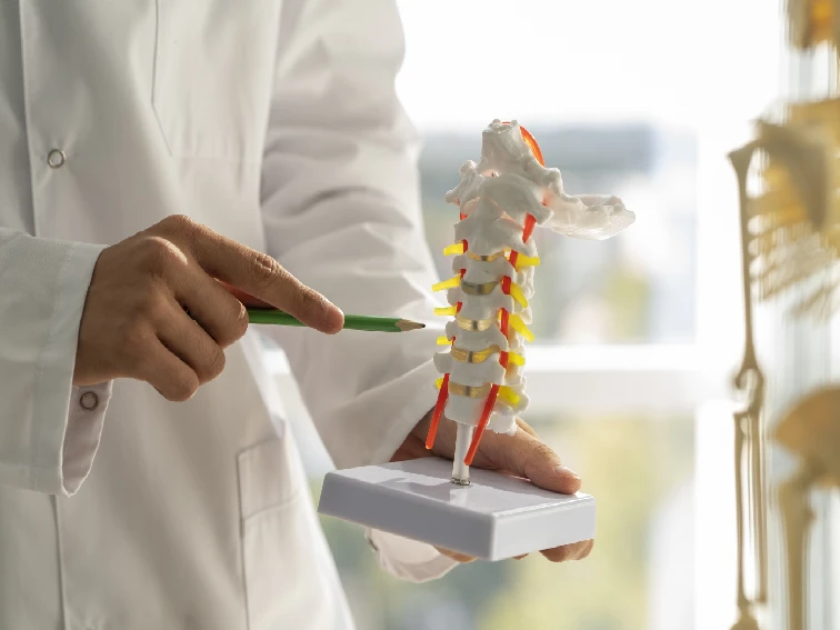
Osteonecrosis: A Silent Threat to Your Bones
Osteonecrosis causes and symptoms: Have you ever wondered how our bones stay strong and healthy? Well, they have a remarkable ability to repair and regenerate themselves. However, sometimes, a condition called Osteonecrosis can silently strike, affecting our bones without warning. In this article, we will delve into the world of Osteonecrosis, exploring its causes, symptoms, treatments, and ways to prevent it. So, let’s embark on this journey to safeguard our precious bones!
1. What is Osteonecrosis?
Osteonecrosis, also known as avascular necrosis, is a medical condition that occurs when a bone loses its blood supply, leading to bone tissue death. Without a proper blood supply, the bone weakens and collapses, causing pain and impaired function. It can affect various bones in the body, including the hips, knees, shoulders, and ankles. Osteonecrosis is a silent threat as it may not show symptoms until significant damage has occurred.
2. Understanding Bone Health – Osteonecrosis causes and symptoms
Bones are like the strong pillars that support our body structure. They are living tissues that undergo a continuous process of renewal. Bone health is vital to ensure overall well-being, as bones protect our vital organs and facilitate movement. Adequate calcium intake, vitamin D, and regular exercise play essential roles in maintaining strong and healthy bones.
3. Unraveling the Osteonecrosis causes and symptoms
While Osteonecrosis can affect anyone, certain risk factors can increase its likelihood. These include:
• Trauma: Injury to bones or joints can disrupt blood flow and lead to Osteonecrosis.
• Medical Conditions: Conditions like sickle cell disease, lupus, and HIV can contribute to bone tissue death.
• Long-term Steroid Use: Prolonged use of steroids can weaken bones and increase the risk of Osteonecrosis.
• Excessive Alcohol Consumption: Heavy alcohol consumption can impair blood flow to bones, leading to Osteonecrosis.
4. Identifying the Symptoms
In the early stages, Osteonecrosis may not cause noticeable symptoms. As the situation advances, you might encounter the following symptoms:
• Pain: Gradual onset of pain that worsens with activity and reduces with rest.
• Stiffness: Restricted movement and stiffness in the affected joint.
• Cracking or Popping Sensation: Sensation of bones rubbing together.
• Limited Mobility: Difficulty in bearing weight on the affected limb.
5. When to Seek Medical Attention
If you experience persistent joint pain or any of the above symptoms, it’s essential to consult a healthcare professional promptly. Early detection can prevent further damage and increase the chances of successful treatment.
6. Diagnosing Osteonecrosis
To diagnose Osteonecrosis, your doctor may perform a thorough physical examination, review your medical history, and order imaging tests like X-rays, MRI, or CT scans. These tests help identify the extent of bone damage and determine the appropriate treatment.
7. Navigating Treatment Options
The choice of treatment depends on the stage of Osteonecrosis and the affected bone. The main goals of treatment are to relieve pain, improve joint function, and prevent further bone damage. Here are some treatment options:
– Medications: Nonsteroidal anti-inflammatory drugs (NSAIDs) can be beneficial in alleviating pain and reducing inflammation. – Physical Therapy: It can enhance joint mobility and strengthen surrounding muscles. – Assistive Devices: Using crutches or braces can relieve pressure on the affected bone. – Core Decompression: This surgical procedure reduces pressure within the bone, allowing better blood flow.
8. Surgical Interventions for Osteonecrosis causes and symptoms
In advanced stages, surgical intervention may be necessary. Some surgical options include:
– Bone Grafting: Healthy bone tissue is transplanted to replace the damaged bone. – Osteotomy: The bone is reshaped to reduce stress on the affected area. – Joint Replacement: For severe cases, joint replacement surgery may be recommended.
9. Non-Surgical Treatments
If Osteonecrosis is detected in its early stages, non-surgical treatments can be effective. These include lifestyle modifications, pain management, and physical therapy. Regular follow-ups with your orthopedic doctor are essential to monitor progress and adjust the treatment plan if needed.
10. Lifestyle Tips for Bone Health
Preventing Osteonecrosis starts with maintaining strong bones. Here are some lifestyle tips to promote Bone Health:
Balanced Diet: Ensure a diet rich in calcium, vitamin D, and other essential nutrients for bone health. – Regular Exercise: Engage in weight-bearing exercises like walking, dancing, or hiking. – Limit Alcohol and Smoking: Reduce alcohol intake and avoid smoking, which can weaken bones.
11. Preventing Osteonecrosis causes and symptoms
Prevention is better than cure, and there are steps you can take to minimize the risk of Osteonecrosis:
Manage Underlying Conditions: If you have medical conditions that increase Osteonecrosis risk, work with your healthcare provider to manage them effectively. Avoid Prolonged Steroid Use: If prescribed steroids, follow your doctor’s recommendations and avoid long-term use whenever possible. – Take care of your joints: Utilize correct techniques and protective equipment while engaging in physical activities to avoid injuries.
12. Living with Osteonecrosis
Living with Osteonecrosis may present challenges, but with the right treatment and lifestyle adjustments, many individuals can manage the condition effectively. Receiving support from healthcare professionals, family, and friends can have a significant impact on how you cope with the condition.
13. Osteonecrosis and Quality of Life
Osteonecrosis can impact a person’s quality of life, especially if it affects mobility and daily activities. Engaging in physical therapy, adhering to treatment plans, and staying positive can improve overall well-being.
14. Osteonecrosis in Different Age Groups
Osteonecrosis can affect people of all ages, but certain age groups may have specific risk factors. Understanding these factors can help tailor prevention and treatment strategies accordingly.
15. Research and Future Perspectives
Medical research is continuously advancing, and there are ongoing studies to better understand Osteonecrosis and improve treatment options. Staying informed about the latest developments can help individuals make well-informed decisions about their health.
Osteonecrosis causes and symptoms FAQs
What are the early signs of Osteonecrosis?
Early signs include joint pain, stiffness, and limited mobility.
Can Osteonecrosis be cured completely?
In some cases, early detection and appropriate treatment can halt or reverse the progression of Osteonecrosis.
Is Osteonecrosis preventable?
While not all cases are preventable, certain lifestyle changes and risk management can lower the risk of Osteonecrosis.
How does Osteonecrosis affect young adults?
Osteonecrosis in young adults can be associated with trauma or underlying medical conditions.
Can physical therapy help with Osteonecrosis?
Yes, physical therapy can improve joint function and mobility in individuals with Osteonecrosis.
conclusion
understanding Osteonecrosis causes and symptoms are crucial for safeguarding our bone health. By staying informed about its causes, symptoms, and treatment options, we can take proactive steps to protect our bones and lead healthier lives. Remember, early detection and timely medical intervention can make all the difference in managing this silent threat effectively. So, let’s prioritize our bone health and take the necessary steps to keep our bones strong and resilient.
Diaphyseal Femur Fractures, Symptoms & Treatment
Diaphyseal femur fracture, also known as femoral shaft fracture, is a common type of bone fracture that occurs in the long bone of the thigh, known as the femur. These fractures typically happen due to high-energy trauma, such as car accidents or falls from a significant height. Understanding the causes, symptoms, treatment options, and recovery process is crucial for individuals dealing with diaphyseal femur fractures. In this article, we will explore these aspects in detail to provide you with valuable information.
Causes of Diaphyseal Femur Fracture
Diaphyseal femur fractures can occur as a result of various causes. The most common cause is trauma, which can be attributed to high-energy accidents like car crashes, motorcycle accidents, or falls from heights. Sports-related injuries, such as those occurring in contact sports like football or skiing, can also lead to femur fractures. Additionally, individuals with weakened bones due to conditions like osteoporosis or bone tumors may be more susceptible to diaphyseal femur fractures even from minor trauma.
Symptoms of Diaphyseal Femur Fractures
When a diaphyseal femur fracture occurs, there are several symptoms that may manifest. These include:
- Severe pain in the thigh or groin area
- Inability to bear weight or walk
- Swelling and bruising around the affected area
- Visible deformity or angulation of the leg
- Limited range of motion in the hip or knee joint
- Numbness or tingling in the leg, indicating potential nerve damage
If you experience any of these symptoms after a traumatic event or suspect a diaphyseal femur fracture, it is important to seek immediate medical attention.
Treatment Options for Diaphyseal Femur Fractures
The treatment of diaphyseal femur fractures depends on various factors, including the severity of the fracture and the patient’s overall health. Here are some common treatment options:
Non-Surgical Treatment: In some cases, if the fracture is stable and the bones are aligned properly, non-surgical treatment methods may be employed. This typically involves immobilizing the leg with a cast or brace to allow the bones to heal naturally over time. However, this approach is less common for diaphyseal femur fractures due to their typically unstable nature.
Surgical Treatment: Surgery is often the preferred treatment for diaphyseal femur fractures, especially if the fracture is displaced or unstable. Surgical options include:
Intramedullary Nailing: This is the most common surgical technique used for diaphyseal femur fractures. It involves inserting a metal rod into the marrow canal of the femur to stabilize the fracture. The rod is usually secured with screws or locking mechanisms to provide stability during the healing process.
Plate and Screw Fixation: In some cases, a plate and screws may be used to fixate the fractured bone. The plate is attached to the outside of the bone, and screws are inserted to hold the fractured fragments together.
External Fixation: This method involves using external frames and pins to stabilize the fracture. It is typically used in cases where there is significant soft tissue damage or when other methods are not feasible.
Recovery Process for Diaphyseal Femur Fractures
The recovery process for diaphyseal femur fractures can be lengthy and requires patience and adherence to medical guidance. Here are some important points to consider during the recovery phase:
Physical Therapy: Following surgery or immobilization, physical therapy plays a crucial role in the recovery process. A physical therapist will guide you through exercises to improve strength, flexibility, and range of motion in the affected leg. This helps prevent muscle atrophy and promotes proper healing.
Weight-Bearing Progression: Initially, you may need to avoid putting weight on the affected leg. As healing progresses, your healthcare provider will gradually allow partial or full weight-bearing activities. It is important to follow their instructions and not rush the process to prevent complications.
Nutrition and Lifestyle: A balanced diet rich in essential nutrients like calcium, vitamin D, and protein is vital for bone healing. Quitting smoking and reducing alcohol consumption can also aid in the recovery process by promoting better bone health.
Follow-up Care: Regular follow-up appointments with your orthopedic specialist are crucial to monitor the healing progress and make any necessary adjustments to your treatment plan.
Conclusion
Diaphyseal femur fractures are significant injuries that require prompt medical attention and appropriate treatment. By understanding the causes, symptoms, treatment options, and recovery process associated with these fractures, individuals can make informed decisions and actively participate in their own healing journey. If you suspect a diaphyseal femur fracture or experience any symptoms, do not hesitate to consult a healthcare professional for a proper diagnosis and guidance towards recovery. Remember, the road to recovery may be challenging, but with proper care and perseverance, you can regain mobility and return to an active lifestyle.
FAQs
What is a diaphyseal femur fracture?
A diaphyseal femur fracture is a type of bone fracture that occurs in the long bone of the thigh, known as the femur. It involves a break in the middle (diaphysis) of the femur bone.
What causes diaphyseal femur fractures?
Diaphyseal femur fractures are commonly caused by high-energy trauma, such as car accidents, falls from heights, or sports-related injuries. Weakened bones due to conditions like osteoporosis can also increase the risk of these fractures.
What are the symptoms of a diaphyseal femur fracture?
Symptoms of a diaphyseal femur fracture may include severe pain in the thigh or groin area, inability to bear weight or walk, swelling and bruising around the fracture site, visible deformity or angulation of the leg, limited range of motion in the hip or knee joint, and numbness or tingling in the leg.
How are diaphyseal femur fractures treated?
Treatment options for diaphyseal femur fractures depend on various factors. Non-surgical treatment methods, such as casting or bracing, may be used for stable fractures. However, surgery is often necessary for displaced or unstable fractures. Surgical options include intramedullary nailing, plate and screw fixation, or external fixation.
What is the recovery process like for diaphyseal femur fractures?
The recovery process for diaphyseal femur fractures can be lengthy. It typically involves physical therapy to regain strength, flexibility, and range of motion in the affected leg. Weight-bearing progression, nutrition and lifestyle considerations, and regular follow-up care are also important aspects of the recovery process.
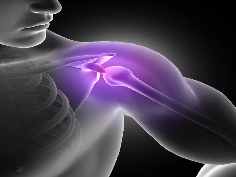
Understanding Shoulder Impingement
Causes, Symptoms, and Treatment
Shoulder impingement syndrome is a common condition that causes pain and discomfort in the shoulder joint. It occurs when the tendons of the rotator cuff muscles become compressed or irritated as they pass through the narrow space between the upper arm bone and the shoulder blade. This article aims to provide a comprehensive understanding of shoulder impingement, including its causes, symptoms, diagnosis, and treatment options.
Causes of Shoulder Impingement Syndrome
Shoulder impingement can have various causes, including
Repetitive Overhead Activities
Engaging in repetitive overhead activities such as throwing, swimming, or weightlifting can lead to shoulder impingement over time. These activities put excessive stress on the rotator cuff tendons, leading to inflammation and irritation.
Poor Posture
Maintaining poor posture, particularly rounded shoulders, can contribute to shoulder impingement. Slumping forward causes the space within the shoulder joint to narrow, increasing the risk of tendon compression.
Muscular Imbalances
Muscular imbalances around the shoulder joint, particularly weak rotator cuff muscles and overactive chest muscles, can disrupt the normal mechanics of the shoulder. This imbalance can result in impingement and pain.
Symptoms and Diagnosis
The symptoms of shoulder impingement may include
Shoulder Pain
Pain is the primary symptom of shoulder impingement. It is often felt on the top and outer side of the shoulder and may worsen with overhead movements.
Limited Range of Motion
Individuals with shoulder impingement may experience a decreased range of motion in their affected shoulder. Activities such as reaching behind the back or lifting objects overhead may become difficult and painful.
Weakness and Instability
Muscle weakness and instability in the shoulder are common symptoms of impingement. This can lead to difficulty in performing everyday tasks that require arm movement.
Diagnosing shoulder impingement typically involves a physical examination, assessment of medical history, and imaging tests such as X-rays, ultrasound, or MRI scans.
Treatment Options for Shoulder Impingement Syndrome
Conservative Treatment: The initial treatment for shoulder impingement often involves conservative measures, including:
Rest and Activity Modification: Avoiding activities that worsen the symptoms and giving the shoulder adequate rest can help alleviate pain and promote healing.
Physical Therapy
A structured physical therapy program can improve shoulder strength, stability, and flexibility. Therapists often use exercises that focus on strengthening the rotator cuff muscles and correcting any muscle imbalances.
Nonsteroidal Anti-Inflammatory Drugs (NSAIDs)
Over-the-counter NSAIDs, such as ibuprofen, can help reduce pain and inflammation associated with shoulder impingement.
Injections
Corticosteroid injections into the shoulder joint may be recommended to provide short-term relief from pain and inflammation.
Surgical Intervention
If conservative treatments do not provide sufficient relief, surgery may be considered. The surgical procedure aims to create more space within the shoulder joint and repair any damaged structures.
Preventing Shoulder Impingement
While shoulder impingement cannot always be prevented, certain measures can help reduce the risk:
Maintain Proper Posture
Maintaining good posture, particularly during activities involving shoulder movement, can help reduce the risk of impingement. Keeping the shoulders back and avoiding slumping forward is crucial.
Warm-up and Stretching
Engaging in proper warm-up exercises and stretching before any physical activity can help prepare the shoulder muscles and reduce the risk of injury.
Strengthen the Rotator Cuff Muscles
Regularly performing exercises that target the rotator cuff muscles can help improve their strength and stability, reducing the likelihood of impingement.
Modify Activities
If you engage in repetitive overhead activities, consider modifying your technique or reducing the frequency or intensity to avoid overloading the shoulder joint.
Bottom Line
Shoulder impingement syndrome can cause significant pain and affect daily activities. Understanding its causes, symptoms, and treatment options is crucial for effective management. Early intervention, including rest, physical therapy, and lifestyle modifications, can often alleviate and promote recovery. If conservative treatments fail, surgical intervention may be necessary. By adopting preventive measures and maintaining shoulder health through exercise and proper posture, individuals can reduce the risk of developing shoulder impingement and enjoy a pain-free and active lifestyle.
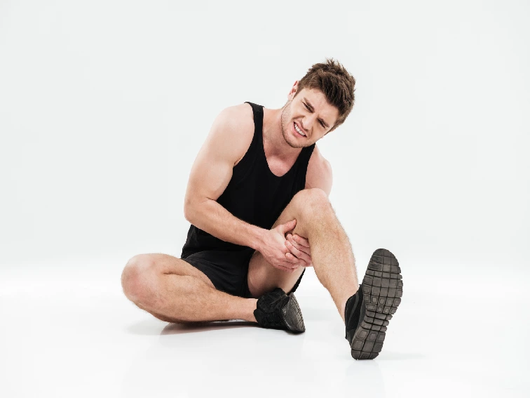
ACL Injury: Causes, Symptoms, Treatment, and Prevention
ACL surgery in Hyderabad | Dr. Praharsha Mulpur
The anterior cruciate ligament (ACL) is a crucial ligament in the knee joint that provides stability and helps in the proper functioning of the knee. However, ACL injuries are quite common, especially among athletes and individuals involved in sports activities. Understanding the causes, symptoms, treatment options, and prevention strategies for ACL injuries is essential for both athletes and the general population. In this article, we will delve into these aspects in detail.
What is an ACL Injury?
An ACL injury refers to the tearing or spraining of the anterior cruciate ligament, which is located in the knee joint. It commonly occurs during sports activities that involve sudden stops, changes in direction, or pivoting movements. ACL injuries can range from mild sprains to complete tears, affecting the stability and mobility of the knee.
Causes of an ACL Injury
ACL injuries can happen due to various reasons, including:
1. Sudden Stops and Direction Changes
Activities that require sudden stops or changes in direction, such as basketball, soccer, and skiing, put immense stress on the knee joint. These abrupt movements can lead to ACL injuries.
2. Landing Improperly
Landing from a jump incorrectly, especially with poor knee alignment, can increase the risk of ACL injury. Athletes who participate in sports like gymnastics and basketball are particularly prone to this type of injury.
3. Direct Impact or Collision
A direct blow or collision to the knee, often seen in contact sports like football and rugby, can cause significant damage to the ACL.
4. Muscle Imbalances and Weakness
Muscle imbalances, especially between the quadriceps and hamstrings, can place excessive strain on the ACL. Weak muscles can lead to poor knee stability, increasing the likelihood of ACL injury.
5. Previous ACL Injury
Having a history of ACL injury increases the risk of re-injury, especially if proper rehabilitation and strengthening exercises were not performed.
Symptoms of ACL Injury
The following symptoms may indicate an ACL injury:
1. Sudden Pain and Swelling
Individuals often experience immediate pain and swelling in the knee following an ACL injury. The swelling may occur within a few hours and can be accompanied by a feeling of instability.
2. Popping Sound or Sensation
At the time of injury, a popping sound or sensation may be felt in the knee joint. This is an indication of a significant ACL tear.
3. Limited Range of Motion
ACL injuries can restrict the range of motion in the knee joint. Individuals may find it challenging to fully straighten or bend their knees.
4. Knee Instability
A feeling of knee instability, as if the knee is giving way or unable to support the body’s weight, is a common symptom of an ACL injury.
5. Difficulty in Walking or Weight-Bearing
ACL injuries can make walking or bearing weight on the affected leg painful and difficult.
Diagnosis of ACL Injury
To diagnose an ACL injury, a healthcare professional will conduct a thorough examination of the knee, review the individual’s medical history, and order additional tests if necessary. These tests may include:
X-rays: X-rays can help rule out any fractures or bone injuries in the knee.
MRI (Magnetic Resonance Imaging): An MRI scan provides detailed images of the knee’s soft tissues, allowing for an accurate assessment of ACL tears or other ligament injuries.
Treatment Options for ACL Injury
The treatment for ACL injury depends on various factors, such as the severity of the injury, the individual’s activity level, and their overall health. The treatment options may include:
1. Surgical Intervention
ACL surgery in Hyderabad: In cases where the ACL is completely torn or if the individual wants to return to high-demand sports, surgical reconstruction of the ligament may be recommended. This procedure involves using a graft to replace the torn ACL.
2. Non-Surgical Treatment
For individuals with partial tears or those who may not require surgery, non-surgical treatment options can be considered. These may include physical therapy, rehabilitation exercises, and bracing to support the knee joint.
Rehabilitation and Recovery
After an ACL injury, a comprehensive rehabilitation program is essential for proper recovery and to regain strength and stability in the knee joint. The rehabilitation process may include:
Physical therapy exercises to improve range of motion, strength, and flexibility.
Balance and proprioception training to enhance knee stability.
Gradual return to sports-specific activities under the guidance of a healthcare professional.
Prevention Strategies for ACL Injury
While it may not be possible to prevent all ACL injuries, several strategies can help reduce the risk:
1. Strengthening Exercises
Regularly engaging in exercises that target the muscles around the knee, such as the quadriceps and hamstrings, can improve knee stability and reduce the risk of ACL injuries.
2. Proper Technique and Landing Mechanics
Learning and practicing proper techniques for sports activities, including correct landing mechanics, can minimize the stress on the knee joint and reduce the chances of ACL injury.
3. Warm-up and Stretching
Performing a thorough warm-up routine and stretching before engaging in sports or physical activities can prepare the muscles and joints for the demands of the activity, reducing the risk of injury.
4. Using Protective Equipment
Wearing appropriate protective equipment, such as knee braces or padding, can provide additional support and reduce the impact on the knee during sports activities.
5. Rest and Recovery
Allowing adequate rest and recovery time between intense training sessions or competitions can help prevent overuse injuries, including ACL injuries.
Returning to Sports After an ACL Injury
Returning to sports after an ACL injury requires careful planning and guidance from healthcare professionals. The process typically involves the following steps:
Assessment and Rehabilitation: The injured individual undergoes a thorough assessment to determine their readiness for sports-specific activities. Rehabilitation exercises and training are tailored to their specific needs.
Gradual Progression: The individual gradually increases the intensity and complexity of their training under the supervision of healthcare professionals, focusing on strength, balance, and agility.
Sports-Specific Training: Once the individual has regained sufficient strength and stability, they can begin sports-specific training to relearn movement patterns and regain confidence.
Return to Competition: The final stage involves a gradual return to competitive play, ensuring the knee is fully rehabilitated and the individual is physically and mentally prepared.
ACL Injury in Children and Adolescents
ACL injuries in children and adolescents require special consideration due to their ongoing growth and development. Treatment approaches may differ, considering factors such as skeletal maturity and the potential impact on future growth.
ACL Injury and Gender Differences
Research suggests that females are more prone to ACL injuries than males. Anatomical, hormonal, and neuromuscular factors may contribute to this gender disparity. Understanding these differences can aid in implementing targeted prevention and training programs.
Long-Term Effects of ACL Injury
While many individuals can recover well from ACL injuries with appropriate treatment and rehabilitation, long-term effects may still be present. These can include a higher risk of developing osteoarthritis in the knee joint later in life.
The Role of Physical Therapy in ACL Injury
Physical therapy plays a crucial role in the treatment and rehabilitation of ACL injuries. Skilled therapists design individualized exercise programs to restore strength, range of motion, and functional abilities.
Surgical Intervention for ACL Injury
ACL surgery in Hyderabad: Surgical intervention for ACL injury aims to reconstruct the torn ligament using a graft. Advances in surgical techniques and rehabilitation protocols have improved outcomes and helped individuals return to their previous level of activity.
Non-Surgical Treatment for ACL Injury
Non-surgical treatment options for ACL injury may be considered for individuals with partial tears or those with lower activity levels. Physical therapy, exercise programs, and bracing can help manage symptoms and restore knee function.
Bottom Line
ACL injuries are common and can significantly impact an individual’s mobility and quality of life. Understanding the causes, symptoms, treatment options, and prevention strategies is essential. By implementing proper techniques, engaging in strengthening exercises, and seeking appropriate medical care, individuals can reduce the risk of ACL injuries and promote long-term knee health.
FAQs
1. Can ACL injuries heal without ACL surgery in Hyderabad?
In some cases, non-surgical treatment options, such as physical therapy and rehabilitation exercises, can help manage ACL injuries. However, the decision depends on the severity of the injury and the individual’s activity level and goals.
2. How long does it take to recover from ACL surgery?
The recovery time after ACL surgery can vary depending on the individual, the extent of the injury, and the rehabilitation process. It generally takes several months for individuals to regain full strength and stability in the knee.
3. Are ACL injuries more common in certain sports?
ACL injuries are more prevalent in sports that involve sudden stops, changes in direction, and pivoting movements, such as basketball, soccer, skiing, and football.
4. Can ACL injuries be prevented?
While ACL injuries cannot be entirely prevented, implementing preventive measures such as strengthening exercises, proper technique, warm-up routines, and using protective equipment can reduce the risk.
5. Can you return to sports after an ACL injury?
With appropriate treatment, rehabilitation, and guidance from healthcare professionals, many individuals can successfully return to sports after an ACL injury. The process involves a gradual progression of training and sports-specific exercises.
To know more about ACL surgery in Hyderabad at KIMS hospitals, meet Dr. Praharsha Mulpur.

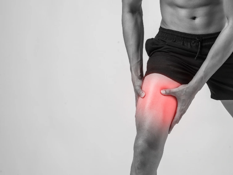
Stress Fractures – Causes, Symptoms & Treatment
Bone fracture treatment in Hyderabad: A stress fracture is a small crack or severe bruising within a bone, typically caused by repetitive stress or overuse. It commonly occurs in weight-bearing bones such as the tibia (shinbone), metatarsals (bones of the foot), or fibula (calf bone), but can also happen in other bones. Athletes, particularly those involved in activities that involve repetitive impact or excessive loading on the bones, are more susceptible to stress fractures. However, anyone can develop a stress fracture under certain circumstances such as: undergoing excessive training without taking enough rest. Starting a new physical activity or sport without appropriate guidance, training or equipment. Spontaneously increasing activity level; training or working out without a proper equipment; playing the same sport without a break between seasons.
What causes Stress Fractures?
The main cause of stress fractures is the repetitive application of force or impact on a bone that does not have adequate time to heal and recover. This can result from activities such as running, jumping, dancing, or participating in sports that involve a lot of running or sudden changes in direction. Factors that increase the risk of stress fractures include sudden increases in activity level, improper training techniques, poor footwear, hard training surfaces, and certain medical conditions that weaken the bones, such as osteoporosis.
What are the risk factors for stress fractures?
The following factors can increase the risk of stress fractures:
Eating disorders, obesity or excess body weight, vitamin D deficiency, flat feet, high-arch feet, osteoporosis and bunions.
Symptoms of Stress Fractures?
The symptoms of a stress fracture may include localized pain that worsens with activity and improves with rest, swelling, tenderness at the site of the fracture, and possible bruising. In some cases, the pain may be present even during rest if the fracture is severe. Other symptoms may include noticeable pain while resting; tenderness in the affected bone even to a light touch. Stress fractures require proper diagnosis and treatment to prevent complications and promote healing.
Diagnosis
If you suspect a stress fracture, it is important to consult an orthopedic doctor. The orthopedic doctor will evaluate your symptoms, medical history, and may order imaging tests such as X-rays, bone scans, or magnetic resonance imaging (MRI) to confirm the diagnosis.
Bone Fracture Treatment in Hyderabad
Treatment typically involves rest, avoiding activities that exacerbate the pain, and protecting the affected area with braces, crutches, or casts if necessary. To manage pain, your orthopedic doctor may recommend over-the-counter pain relievers or prescribed medications. Physical therapy and rehabilitation exercises are often prescribed to strengthen the surrounding muscles and help prevent future injuries.
Elevating the affected bone: If you have stress fracture in your foot or leg then keep the injured bone above the level of your heart by propping your leg up with cushions and pillows while laying down.
Surgery
Your orthopedic doctor may suggest surgery if your stress fracture doesn’t heal as it should or when you have severe symptoms.
Prevention of stress fractures
Prevention is crucial when it comes to stress fractures. Some preventive measures include gradually increasing activity levels, using proper footwear that provides support and cushioning, maintaining a balanced diet rich in calcium and vitamin D for bone health, cross-training to vary the types of activities and reduce repetitive stress on specific bones, and listening to your body by taking breaks and allowing for proper rest and recovery.
Bottom Line
Pain is often the first symptom of a stress fracture. First, stop the activity that is causing pain. It is important to consult an orthopedic doctor for a proper diagnosis and treatment plan if you suspect a stress fracture or have any concerns about your bone health.