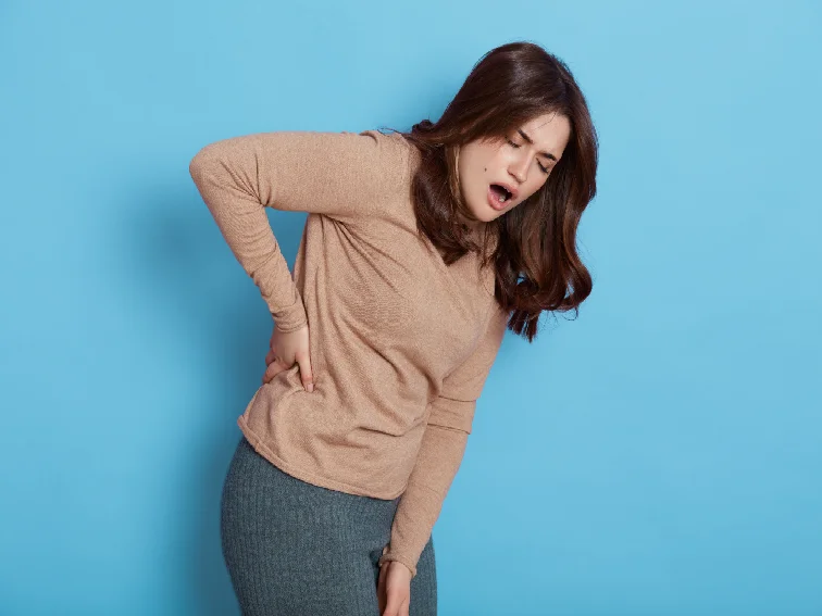
Knee Pain Due to Back Problems: Understanding the Connection
Knee pain is a common ailment that affects people of all ages and lifestyles. While it’s often attributed to issues within the knee joint itself, there’s a less known but crucial connection between knee pain and problems in the lower back. In this article, we will explore how back problems can lead to knee pain and what you can do to alleviate it.
The Anatomy of the Connection
To understand the relationship between knee pain and back problems, it’s essential to grasp the interconnected nature of our musculoskeletal system. Our body is a complex network of bones, muscles, and joints, all working in harmony to facilitate movement. The lower back, in particular, plays a pivotal role as it supports the upper body and connects it to the lower limbs.
The lumbar spine, located in the lower back, is comprised of five vertebrae. These vertebrae are connected by intervertebral discs, which act as shock absorbers and allow for flexibility. Nerves from the spinal cord branch out from the lumbar region, traveling down to various parts of the body, including the legs and knees.
Knee Pain due to Back Problems
1. Nerve Compression: One of the primary ways that back problems can lead to knee pain is through nerve compression. When the spinal nerves in the lower back become compressed or irritated due to issues like herniated discs or spinal stenosis, the pain can radiate down the legs and affect the knees. This referred pain can be intense and is often mistaken for primary knee issues.
2. Altered Gait and Posture: Back problems can also result in changes to your gait and posture. When you experience pain or discomfort in your lower back, you may unconsciously alter the way you walk or stand. These adjustments can put extra stress on your knee joints, leading to pain over time.
3. Muscle Imbalances: Back problems can disrupt the delicate balance of muscles that support the spine and pelvis. When this balance is disrupted, it can lead to compensatory movements that strain the knee joints. For example, weak or tight muscles in the lower back and hip area can affect the alignment of the knees.
Common Back Problems Linked to Knee Pain
Several back problems are commonly associated with knee pain:
1. Herniated Discs: When the soft inner material of a spinal disc protrudes through its outer shell, it can press on nearby nerves, causing pain that radiates down to the knees.
2. Spinal Stenosis: This condition involves the narrowing of the spinal canal, which can compress spinal nerves and result in knee pain.
3. Sacroiliac Joint Dysfunction: Dysfunction in the sacroiliac joint, located at the base of the spine, can cause pain that is felt in the lower back and can also radiate down to the knees.
4. Muscle Imbalances: As mentioned earlier, imbalances in the muscles that support the spine and pelvis can contribute to knee pain.
Seeking Relief
If you’re experiencing knee pain due to back problems, it’s crucial to seek professional medical advice. A healthcare provider can conduct a thorough evaluation, which may include imaging tests, to pinpoint the root cause of your pain. Treatment options may include physical therapy, pain management techniques, exercises to strengthen core and leg muscles, and in some cases, surgery.
Bottom Line
knee pain can be a symptom of underlying back problems, emphasizing the importance of a holistic approach to healthcare. Understanding the interconnected nature of our musculoskeletal system and seeking prompt medical attention when experiencing pain can lead to effective treatment and a better quality of life. Remember, addressing the root cause is key to finding lasting relief from knee pain due to back problems.

Capitellar Fractures Symptoms, Causes, and Treatment
Capitellar fractures, though relatively uncommon, can have a significant impact on a person’s life. These fractures, often caused by traumatic incidents, affect the rounded end of the humerus bone in the elbow joint. In this comprehensive article, we’ll delve into the various aspects of capitellar fractures, from their causes and symptoms to the available treatment options.
Let’s explore this topic in detail, ensuring you have all the information you need about capitellar fractures.
Capitellar Fractures Symptoms and Types
Capitellar fractures refer to injuries to the rounded head of the humerus bone, which forms part of the elbow joint. They are typically classified into three main types:
Type I – Non-displaced Fractures
In non-displaced fractures, the broken bone fragments remain in their original position. This type of fracture is less severe and painful and often requires conservative treatment.
Type II – Partially Displaced Fractures
Partially displaced fractures involve a shift in the position of some bone fragments. These fractures may require more advanced treatment approaches.
Type III – Displaced Fractures
Displaced fractures occur when the bone fragments separate significantly. These are the most severe and painful. They are complex capitellar fractures, often necessitating surgical intervention.
Causes of Capitellar Fractures
Understanding the causes of capitellar fractures is crucial for prevention. These fractures are most commonly associated with:
Trauma: High-impact injuries, such as falls, sports accidents, or automobile collisions, are often responsible for capitellar fractures.
Direct Blows: A direct blow to the elbow, especially when the arm is extended, can lead to this type of fracture.
Overuse: Chronic overuse of the elbow joint, common in sports like gymnastics and baseball, can weaken the bone and increase the risk of fractures.
Capitellum fracture symptoms
Identifying the symptoms of a capitellar fracture is essential for prompt diagnosis and treatment:
Pain: Severe pain in the elbow, which worsens with movement, is a common symptom.
Swelling: Swelling around the elbow joint may be noticeable.
Limited Range of Motion: Difficulty in bending or straightening the elbow is a key indicator.
Tenderness: The affected area is tender to the touch.
Diagnosis and Treatment Options
Proper diagnosis is crucial in determining the appropriate treatment. It typically involves:
Imaging: X-rays and sometimes CT scans help in assessing the extent of the fracture.
Non-Surgical Treatment
For non-displaced or some partially displaced fractures, non-surgical approaches may be effective:
Immobilization: Wearing a cast or brace to immobilize the elbow allows the bone to heal.
Physical Therapy: Rehabilitation exercises can aid in regaining strength and range of motion.
Surgical Intervention
For more severe fractures, surgery is often necessary
Open Reduction and Internal Fixation (ORIF): Surgeons realign and stabilize the bone fragments using pins, screws, or plates.
Joint Replacement: In extreme cases, joint replacement may be considered.
FAQs (Frequently Asked Questions)
Can capitellar fractures heal without surgery?
Yes, non-displaced fractures can often heal with non-surgical treatment like casting and physical therapy.
How long does the recovery process take after surgery?
Recovery time varies, but it can take several weeks to months to regain full strength and function.
Are there any long-term complications associated with capitellar fractures?
While most patients recover well, some may experience reduced range of motion or arthritis in the elbow over time.
Can children experience capitellar fractures?
Yes, children involved in sports or accidents can sustain these fractures, and their treatment may differ from adults.
Is there any way to prevent capitellar fractures?
Wearing protective gear during sports and avoiding risky behaviors can reduce the risk of such fractures.
Are there any advancements in the treatment of capitellar fractures?
Ongoing research is exploring new surgical techniques and materials for more effective treatment.
Bottom Line
Capitellar fractures can be challenging, but with the right diagnosis and treatment, many individuals can regain full function of their elbow joint. Remember to seek prompt medical attention if you suspect a capitellar fracture as early intervention can lead to better outcomes.
We hope this article has provided you with valuable insights into the causes, symptoms, and treatment options for capitellar fractures. Stay informed, stay safe, and prioritize your elbow health.

Bone Fracture Repair – What You Must Know
A fractured bone or broken bone is a common injury that occurs when there is a break or crack in a bone. A bone fracture can be partial or complete – and can range from hairline fracture to severe and complex fractures. Treatment for a fractured bone depends on the type, location and severity of the fracture. To fix bone fractures, orthopedic doctors use a wide range of techniques and procedures including uses of braces, casting, splinting and surgical fixation. Surgical repair of fractures depends on the type, location and severity of the fracture.
To ensure the best outcomes for a patient, proper follow up care and healing process are essential following the treatment.
Types of Bone Fractures
Closed Fracture: The bone is broken but does not penetrate the skin.
Open Fracture: The bone breaks through the skin, increasing the risk of infection.
Displaced Fracture: The bone fragments are not aligned properly.
Non-Displaced Fracture: The bone fragments remain in their normal alignment.
Comminuted Fracture: The bone shatters into multiple pieces.
Greenstick Fracture: In this type of fracture, the bone bends and cracks but does not completely break. The fracture doesn’t extend all through the bone. It is common in children.
Stress Fracture: A hairline crack due to repetitive stress, often seen in athletes.
Immediate First Aid
If you suspect a bone fracture, take the following steps:
Immobilize the Injury: Keep the injured area as still as possible to prevent further damage.
Elevate: If applicable, elevate the injured limb to reduce swelling.
Apply Ice: Place an ice pack (wrapped in a cloth) on the injured area for 15-20 minutes.
Seek Medical Help: Fix an appointment with an orthopedic doctor or visit the nearest emergency room for professional evaluation and treatment.
Why you need a specialist doctor to fix your fractured bone?
A specialist orthopedic doctor has the expertise to restore the normal anatomy of the fractured bone.
It is better to consult an orthopedic doctor for fractured bone repair to prevent future complications.
Untreated fractures can undergo unusual healing and become weak and lose function.
If you don’t get proper professional care, you would face the following problems:
- The fractured bone might not heal properly (non-union fracture).
- The bone may heal in an unusual position (malunion fracture).
- The fracture might take longer time than usual to heal (delayed union fracture).
- The other complications of an untreated fractured bone may include infections at the fracture site and formation of blood clots.
Medical Assessment
An orthopedic doctor will assess the fracture through physical examination and imaging tests (X-rays, CT scans, or MRIs) to determine the type and severity of the fracture.
Treatment Options
Casting: Non-displaced fractures can often be treated with a cast to immobilize the bone, allowing it to heal naturally. Your doctor repositions and immobilizes your broken bone with a plaster of Paris or fiberglass cast. The cast holds the broken bone and allows it to heal in a correct position.
Splinting: Used for initial immobilization, especially in open fractures, until further treatment can be administered.
Surgery: Required for complex fractures, open fractures, or those that cannot be realigned through non-invasive methods. Surgical options include:
Internal Fixation: Orthopedic doctors use screws, plates, or rods to stabilize the bone.
External Fixation: A frame placed outside the body to hold the bone in place.
Bone Grafting: It involves transplanting bone tissue to help in fixing the fracture and healing.
Medication: Pain management and antibiotics (for open fractures) may be prescribed.
Rehabilitation
Rehabilitation plays a crucial role in the recovery process:
Physical Therapy: Helps in restoring strength, flexibility, and a range of motions in the affected area.
Weight-Bearing Progression: This involves gradual return to normal activities as guided by a healthcare professional.
Follow-up Care
Regular follow-up appointments are crucial to monitor the healing progress, adjust treatment plans, and address any issues that may arise.
Prevention of Fractures
Bone fractures can lead to complications such as infection, delayed healing, or malunion (improper bone alignment). To prevent fractures, especially in high-risk individuals:
- Maintain a balanced diet rich in calcium and vitamin D.
- Engage in weight-bearing exercises to strengthen bones.
- Use proper protective gear during physical activities.
- Create a safe home environment to prevent falls.
Conclusion
Bone fracture repair involves a combination of immediate first aid, medical assessment, and appropriate treatment options. With proper care, most fractures can heal effectively, allowing patients to regain full functionality in the affected area. It’s essential to follow medical advice and engage in rehabilitation for a successful recovery.
If you are looking for immediate care for a fractured bone, then meet us…
Understanding and Treating Rotator Cuff Tears
Rotator cuff surgery in Hyderabad
Welcome to our comprehensive guide on understanding and effectively treating rotator cuff tears. As a trusted source of information, we are committed to providing you with accurate and detailed insights into this common shoulder injury.
Introduction
Rotator cuff: A group of four muscles that form as tendons and cover around (envelop) the humerus head and stabilize the shoulder joint. They attach the humerus to the shoulder blade. They keep your Shoulder in its socket and help to rotate and lift your arm.
A tear in any of these tendons can result in pain, limited mobility, and reduced quality of life.
Rotator cuff tears are a prevalent medical condition that affects a significant number of individuals, particularly those engaged in repetitive overhead activities or heavy lifting.
Types of Rotator Cuff Tears
Partial Thickness Tears: These tears affect only a portion of the tendon, often causing mild discomfort and limited impact on daily activities. Partial tears do not detach the tendon from the bone.
Full Thickness Tears: These tears extend through the entire tendon, leading to more significant pain as there is a detachment of a part of the tendon. Reduced range of motion, and potential weakness in the affected arm are the common symptoms.
Causes and Risk Factors
Several factors can contribute to the development of rotator cuff tears:
Age: The risk of tears increases with age as tendons naturally degenerate over time.
Overuse: Repetitive overhead motions, common in sports like tennis or baseball, can lead to tears.
Trauma: A sudden fall or impact can result in an acute tear.
When you lift any heavy object with a jerking motion or fall down on your outstretched arm, your rotator cuff can tear. If you fall down and have wrist fracture, a dislocated shoulder or a broken collarbone, then you can have rotator cuff tear.
Poor blood supply to the tendons and repetitive stress or bone spurs can make tears more likely.
Common symptoms include
Pain: Often felt at the front or side of the shoulder, which may radiate down the arm.
Weakness: Difficulty lifting or rotating the arm, especially when holding objects.
Limited Range of Motion: Difficulty reaching behind the back or overhead.
Night Pain: Discomfort that disturbs sleep, particularly when lying on the affected side.
You can feel pain even at rest and over time pain reliever medications do not offer any help. The weakness and pain do not let you perform routine activities such as combing and reaching behind your back.
Recognizing the symptoms of a rotator cuff tear is crucial for early diagnosis and intervention.
Diagnosis
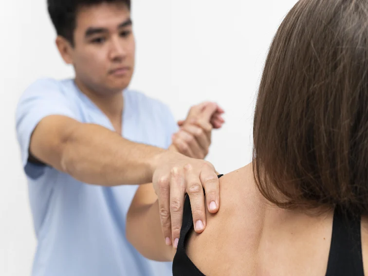
If you suspect a rotator cuff tear, seeking medical attention is vital. An orthopedic doctor will conduct a thorough assessment, which may include:
Physical Examination: Testing the range of motion, strength, and areas of tenderness.
Imaging: X-rays or MRI scans to visualize the tear’s location and severity.
Treatment Options
The main objective of your treatment is to relieve pain and restore function. To ensure this your doctor has several treatment options to choose from. The best treatment approach for a rotator cuff is different for every person. Your orthopedic doctor may consider your overall health, age, activity levels and the type of tear you have.
Conservative Approaches: For mild tears, non-surgical methods may be effective:
Rest and Activity Modification: Allowing the tendon to heal with reduced strain.
Physical Therapy: Strengthening the surrounding muscles can provide support.
Pain Management: Over-the-counter pain relievers and ice packs can help.
Rotator Cuff Surgery in Hyderabad
If non-surgical methods do not offer any help, your doctor may recommend the following surgical interventions for severe tears:
Arthroscopic Repair: Minimally invasive surgery to reattach the tendon.
Open Surgery: Traditional approach for complex tears or reattachments.
Your doctor considers surgical repair if you have a rotator cuff tear due to an acute injury or a recent fall and also when you have significant weakness and loss of function in your shoulder.
Rehabilitation
Regardless of the chosen treatment path, rehabilitation is crucial for restoring shoulder function.
Physical Therapy: Customized exercises to improve strength and flexibility.
Gradual Return to Activities: Controlled reintegration of activities to prevent re-injury.
Prevention
While not all tears can be prevented, certain measures can reduce the risk:
Proper Technique: Learn and practice correct techniques for sports and lifting.
Warm-up and Stretching: Adequate warm-up and stretching before activities.
Cross-Training: Engage in a variety of exercises to avoid overuse of specific muscles.
Bottom Line
In conclusion, understanding the causes, symptoms, and treatment options for rotator cuff tears is essential for effective management and recovery. Whether through conservative methods or surgical interventions, a combination of medical expertise and patient commitment is key to restoring shoulder health and overall well-being.
if you suspect a rotator cuff tear, and looking for rotator cuff surgery in Hyderabad consulting an orthopedic surgeon having expertise in rotator cuff repair is crucial for accurate diagnosis and personalized treatment recommendations.
By providing comprehensive information on rotator cuff tears, we aim to equip you with the knowledge necessary to make informed decisions about your shoulder health. If you have any other concerns, always feel free to contact us.

Tenosynovitis: Understanding Symptoms, Causes & Treatment
Tenosynovitis is a condition that involves the inflammation of the synovium, a thin layer of tissue that covers the tendons in your body. This inflammation can cause pain, swelling, and discomfort, often affecting your ability to move the affected joint. In this comprehensive guide, we’ll delve into the details of Tenosynovitis, including its symptoms, causes, treatment options, and preventive measures.
Tenosynovitis Causes
Tenosynovitis occurs when the synovium, which is responsible for lubricating and protecting tendons, becomes inflamed due to various factors. It most commonly affects the tendons in the wrists, hands, and feet. This condition can result from overuse, repetitive movements, injury, or certain underlying medical conditions.
Tenosynovitis: Understanding the Symptoms
Tenosynovitis is characterized by several noticeable symptoms that can vary in severity. Some of the common symptoms include:
Pain and Tenderness: Individuals with Tenosynovitis often experience localized pain and tenderness around the affected tendon. The pain might worsen with movement or pressure.
Swelling and Redness: Inflammation of the synovium can lead to swelling and redness in the affected area.
Limited Range of Motion: Due to the inflammation and swelling, the affected joint may become stiff, limiting your ability to move it comfortably.
Crepitus: Some people might notice a cracking or creaking sensation when moving the affected joint.
What Causes Tenosynovitis?
Tenosynovitis Causes: Tenosynovitis can be caused by various factors, including:
Repetitive Movements: Engaging in repetitive motions, such as typing, playing musical instruments, or sports activities, can strain the tendons and lead to inflammation.
Injury or Trauma: Direct injury to the tendons, such as sprains or fractures, can trigger an inflammatory response in the synovium.
Medical Conditions: Certain conditions like rheumatoid arthritis or gout can increase the risk of developing Tenosynovitis.
Bacterial Infections: In some cases, bacterial infections can lead to the inflammation of the synovium.
Effective Treatment Options
Treating Tenosynovitis involves a combination of strategies aimed at reducing inflammation and relieving pain. Some approaches include:
Rest and Immobilization: Giving the affected tendon time to rest and heal is essential. Immobilization through splints or braces can aid in the healing process.
Medications: Nonsteroidal anti-inflammatory drugs (NSAIDs) can help reduce pain and inflammation. In severe cases, corticosteroid injections may be recommended.
Physical Therapy: Physical therapy can help improve joint mobility, strengthen surrounding muscles, and prevent recurrences.
RICE Method: Rest, Ice, Compression, and Elevation can help alleviate symptoms and promote healing.
Preventive Measures for Tenosynovitis
Preventing Tenosynovitis involves adopting healthy habits and making ergonomic adjustments:
Proper Ergonomics: Maintain proper posture and ergonomic practices, especially during repetitive tasks, to reduce strain on tendons.
Regular Breaks: Take frequent breaks during activities that involve repetitive motions to give your tendons time to rest.
Warm-Up and Stretching: Prior to engaging in physical activities, warm-up exercises and stretching can help prepare your tendons and muscles.
Balanced Diet: Consuming a diet rich in nutrients can support overall joint health and reduce the risk of inflammation.
FAQs about Tenosynovitis Causes
Q: Can Tenosynovitis occur suddenly?
A: Yes, Tenosynovitis can develop suddenly, especially after overuse or injury.
Q: Is Tenosynovitis a chronic condition?
A: It can be chronic if not properly managed, but with appropriate treatment, many cases resolve within weeks.
Q: Are there any risk factors for developing Tenosynovitis?
A: Yes, factors like repetitive motions, certain medical conditions, and infections can increase the risk.
Q: Can Tenosynovitis affect any joint in the body?
A: While it can occur in various joints, it most commonly affects wrists, hands, and feet.
Q: Is surgery always required for Tenosynovitis?
A: Surgery is usually considered after conservative treatments have failed to provide relief.
Q: Can Tenosynovitis recur after successful treatment?
A: Yes, if preventive measures aren’t taken, there’s a chance of recurrence.
Conclusion
Tenosynovitis can be a challenging condition to deal with, but with the right approach to treatment and prevention, its impact can be minimized. By understanding tenosynovitis causes, symptoms, and effective management strategies, individuals can regain comfort and mobility in their daily lives.