Snapping Hip Syndrome: Causes, Symptoms, and Treatment
Here is your passage with natural transition words added to improve the Yoast transition-word score while keeping the original meaning and structure:
When it comes to hip-related issues, one condition that often perplexes individuals is Snapping Hip Syndrome. Therefore, this article aims to shed light on the condition, its causes, symptoms, diagnosis, treatment options, and prevention techniques. In addition, understanding the intricacies of Snapping Hip Syndrome can help individuals take appropriate steps to manage its impact on their daily lives.
Understanding Snapping Hip Syndrome
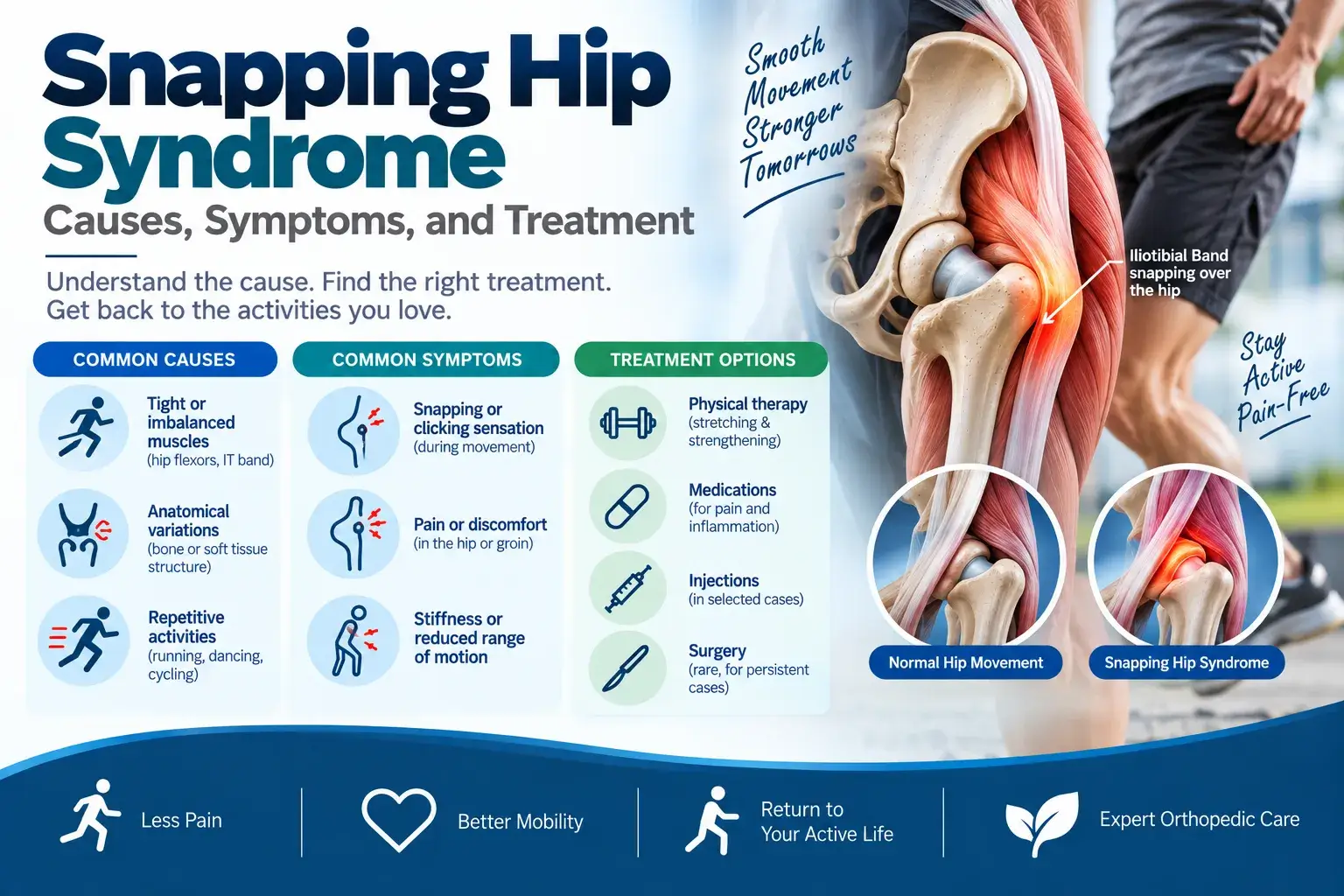
Snapping Hip Syndrome, also known as coxa saltans or dancer’s hip, is a condition characterized by a snapping sensation or sound in the hip joint during certain movements. In particular, it occurs when a muscle, tendon, or ligament moves across a bony prominence in the hip joint. As a result, this movement causes a snapping sensation that may be audible or palpable.
For example, people with this syndrome may hear a snapping sound when they get up from a chair or bed, walk, run, or move their legs. In most cases, the main symptom involves a snapping sound or sensation during movement. However, performers such as sportspersons, athletes, and dancers may also experience pain and weakness that can interfere with their performance.
Definition and Causes
To understand the condition more clearly, doctors classify Snapping Hip Syndrome into three main categories based on its causes: internal, external, and intra-articular. First, internal snapping occurs when the iliopsoas tendon moves over the iliopectineal eminence or femoral head. In contrast, external snapping occurs when the iliotibial band slides over the greater trochanter. Finally, intra-articular snapping involves structures within the hip joint, such as loose bodies or labral tears.
2. Types of Snapping Hip Syndrome
There are two primary types of Snapping Hip Syndrome: extra-articular and intra-articular. Extra-articular snapping is more common and typically caused by the iliotibial band or the iliopsoas tendon. Intra-articular snapping is less prevalent and often associated with structural abnormalities within the joint.
Symptoms and Diagnosis
3.1 Audible Snapping
One of the hallmark symptoms of Snapping Hip Syndrome is an audible snapping sound. This sound may occur during certain movements, such as walking, running, or getting up from a chair. The snapping can be accompanied by a visible movement or jerk in the hip.
3.2 Pain and Discomfort
Here is the active-voice version, with the passive constructions removed:
In addition to the audible snapping, individuals with Snapping Hip Syndrome may experience pain or discomfort. This pain can range from mild to severe and may affect the hip joint or spread to other areas, such as the thigh or buttock.
3.3 Physical Examination and Imaging
To diagnose Snapping Hip Syndrome, a healthcare professional will conduct a physical examination and ask about the patient’s medical history. The healthcare professional may also use imaging techniques, such as X-rays, ultrasound, or magnetic resonance imaging (MRI), to identify structural abnormalities or rule out other conditions.
Treatment Options
4.1 Conservative Approaches
In most cases, conservative treatment approaches are sufficient to manage Snapping Hip Syndrome. This may involve rest, activity modification, applying ice, and the use of non-steroidal anti-inflammatory drugs (NSAIDs) to reduce pain and inflammation.
4.2 Physical Therapy
Physical therapy plays a crucial role in the treatment of Snapping Hip Syndrome. Therapeutic exercises, such as stretching and strengthening routines, can improve flexibility, stability, and muscle imbalances around the hip joint. Manual therapy techniques, such as massage and joint mobilization, may also be employed to alleviate symptoms.
4.3 Surgical Intervention
In rare cases where conservative treatments fail to provide relief, surgical intervention may be considered. The type of surgery depends on the underlying cause of the snapping and may involve releasing or repairing the affected structures.
In hip arthroscopy, an orthopedic surgeon uses small and thin instruments ( arthroscope) by making small incisions. The procedure is most often performed to repair fragments of a torn labrum.
Prevention and Management
5.1 Warm-up and Stretching
To prevent or manage Snapping Hip Syndrome, individuals should engage in a proper warm-up routine before physical activities. Dynamic stretching exercises specific to the hip joint can help improve flexibility and reduce the risk of snapping.
5.2 Strengthening Exercises
Strengthening exercises focusing on the hip muscles can enhance joint stability and prevent excessive movement that may contribute to snapping. These exercises may include hip abductor, adductor, and gluteal strengthening routines.
5.3 Rest and Recovery
Allowing the hip joint sufficient time to rest and recover between activities is crucial. Overuse or repetitive movements can exacerbate snapping symptoms, so individuals should listen to their bodies and avoid activities that cause discomfort.
Conclusion
Snapping Hip Syndrome can be a bothersome condition, but with the right understanding and management strategies, individuals can regain control over their hip health. By adopting a comprehensive approach that includes conservative treatments, physical therapy, and preventive measures, individuals can reduce pain, improve functionality, and enjoy an active lifestyle.
FAQs
- Can Snapping Hip Syndrome go away on its own?
While some cases of Snapping Hip Syndrome may resolve on their own with rest and activity modification, others may require treatment and intervention.
- Is Snapping Hip Syndrome more common in athletes?
Snapping Hip Syndrome can occur in individuals of all activity levels, including athletes, dancers, and sedentary individuals.
- Can Snapping Hip Syndrome lead to long-term complications?
If left untreated or ignored, Snapping Hip Syndrome may lead to chronic pain, hip joint instability, or the development of other hip conditions.
- Are there any exercises that can worsen Snapping Hip Syndrome?
Certain exercises that involve repetitive hip movements or excessive stress on the joint may worsen snapping symptoms. It is best to consult with a healthcare professional or physical therapist for personalized guidance.
- Can Snapping Hip Syndrome be prevented?
While not always preventable, adopting proper warm-up routines, engaging in strengthening exercises, and allowing adequate rest and recovery can help reduce the risk of developing Snapping Hip Syndrome.
Ganglion cysts – Causes, Diagnosis & Treatment
Ganglion cyst treatment in Hyderabad – Dr. Praharsha Mulpur
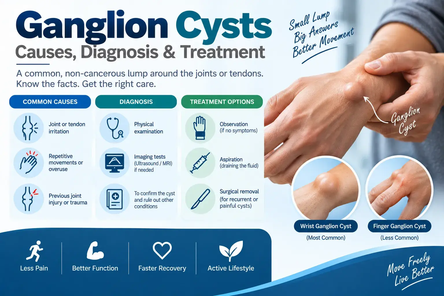
manifest as masses that tend to emerge along the tendons or joints, primarily on the wrists or hands. It is also possible for them to develop in the ankles and feet. Ganglion cysts are usually spherical or elliptical in shape and are filled with a jellylike substance. They are not tumors
Ganglion cysts of a small size can resemble a pea. The size can alter. If a nearby nerve gets pressed, ganglion cysts can cause pain. Occasionally, they impact the mobility of the joints.
Features of Ganglion Cysts
Ganglion cysts typically exhibit these characteristics:
Location-The common location where ganglion cysts form is along the tendons or joints situated on the wrists or hands. Ankles and feet are next areas where ganglion cysts may form. Cysts can also develop in close proximity to other joints.
The shape of ganglion cyst is either circular or elliptical. Some cysts are so small to sense. Cysts tend to grow in size over time due to movements in the joint.
Pain-Typically, ganglion cysts do not cause any pain. In case a cyst applies pressure on a nerve or any other structures, it may result in muscle weakness, numbness, tingling, or pain.
When to See a doctor?
If you detect a lump or experience discomfort in your wrist, hand, ankle, or foot, it is advisable to consult an orthopedic doctor. The doctor will make a diagnosis to help assist whether management is necessary.
Causes
The reason behind a ganglion cyst remains unknown. A small water balloon-like structure on a stem originates from a tendon’s lining or joint to form the cyst. The cyst contains a viscous liquid that is similar to the one found in the joints or the areas surrounding tendons.
What are the risk factors for Ganglion Cysts?
Factors that can raise the likelihood of developing a ganglion cyst include:
Age and gender– Although anyone is susceptible to developing ganglion cysts, women aged 20 to 40 years may most often have them.
Osteoarthritis- Individuals with degenerative arthritis in the finger joints located closest to the nail beds have an increased likelihood of developing ganglion cysts in close proximity to those joints.
Tendon or joint injury– An injury to a tendon or joint increases the likelihood of developing a ganglion cyst.
Diagnosis
Your orthopedic doctor, during a physical examination, may apply pressure on the cyst to determine if it causes pain. To determine whether the cyst is solid or filled with fluid, your doctor shines it by passing a light through it. To confirm the diagnosis and exclude other conditions like arthritis or a tumor, your doctor orders imaging tests such as X-rays, ultrasounds, or MRIs. Confirming the diagnosis is also possible by extracting fluid from the cyst with a needle. Ganglion cyst fluid is usually clear and dense.
Ganglion Cyst Treatment in Hyderabad
If you have a painless ganglion cyst, with no other symptoms, your doctor may choose an observational approach where they monitor it and do not treat it unless necessary. Often, the cysts grow and shrink. Some go away on their own.
A ganglion cyst can grow in size with activity. Wearing a Brace or Splint can help because, they keep the joint still. It might help for a time to shrink the cyst and relieve pain and pressure on nerves. However, long-term use can weaken surrounding nerves and muscles.
If the cyst is hindering the mobility of your joint treatment is necessary. For a ganglion cyst that causes problems, an orthopedic doctor drains the cyst with a needle. If the cyst is causing severe problems, then it is removed surgically.
For the best ganglion cyst treatment, meet Dr. Praharsha Mulpur
Achilles Tendinitis Types, Symptoms & Treatment
Achilles tendinitis is a common problem that occurs due to inflammation and irritation of the large tendon that runs down the back of the lower leg. This tendon, connects the calf muscles to the heel bone. It is responsible for facilitating several movements, including walking, running, climbing stairs, jumping, and standing on tiptoes.
Here is the active-voice version with the passive constructions removed: Despite its ability to withstand significant stress from these activities, overuse can lead to tendinitis, a common condition that affects the Achilles tendon.
Overuse can cause Achilles tendon tendinitis, which affects the strong band of tissue that connects the calf muscles to the heel bone in the lower leg. Runners often develop this injury after suddenly increasing the intensity or duration of their runs. Middle-aged individuals who partihttps://chatgpt.com/how-to-keep-your-joints-healthy/cipate in weekend sports such as basketball or tennis can also develop Achilles tendinitis.
This removes passive constructions such as “It is caused by” and “is typically seen in runners.”
Description
Another term you might encounter is “tendinopathy,”. It describes a condition where the tendon experiences microscopic degeneration due to extensive damage over time. Tendinitis, tendinosis, and tendinopathy are interchangeable terms that describe the same issue.
Achilles Tendonitis Vs Achilles Rupture
Use this active-voice version: Achilles tendinitis differs from an Achilles rupture. An Achilles rupture occurs when a sudden injury tears the tendon in half or separates it from the heel bone. Sudden injuries often cause Achilles ruptures.
Use this active-voice version: This wording removes the passive construction “is often caused by” and keeps the explanation clear and direct.
The primary subject of discussion of this blog will be Achilles tendinitis rather than Achilles tendon rupture.
Symptoms of Achilles Tendinitis
- Achilles tendinitis causes
- Pain in the back of the heel (while wearing shoes)
- Persistent swelling in the affected area
- Formation of bone spur (insertional tendinitis)
- Thickening of the tendon
- Chronic pain after exercise
- Pain along the back of the heel or tendon that worsens with activity
- Pain and stiffness in the Achilles tendon
Achilles Tendinitis Types
There are two types –
- Nonintentional Achilles tendinitis
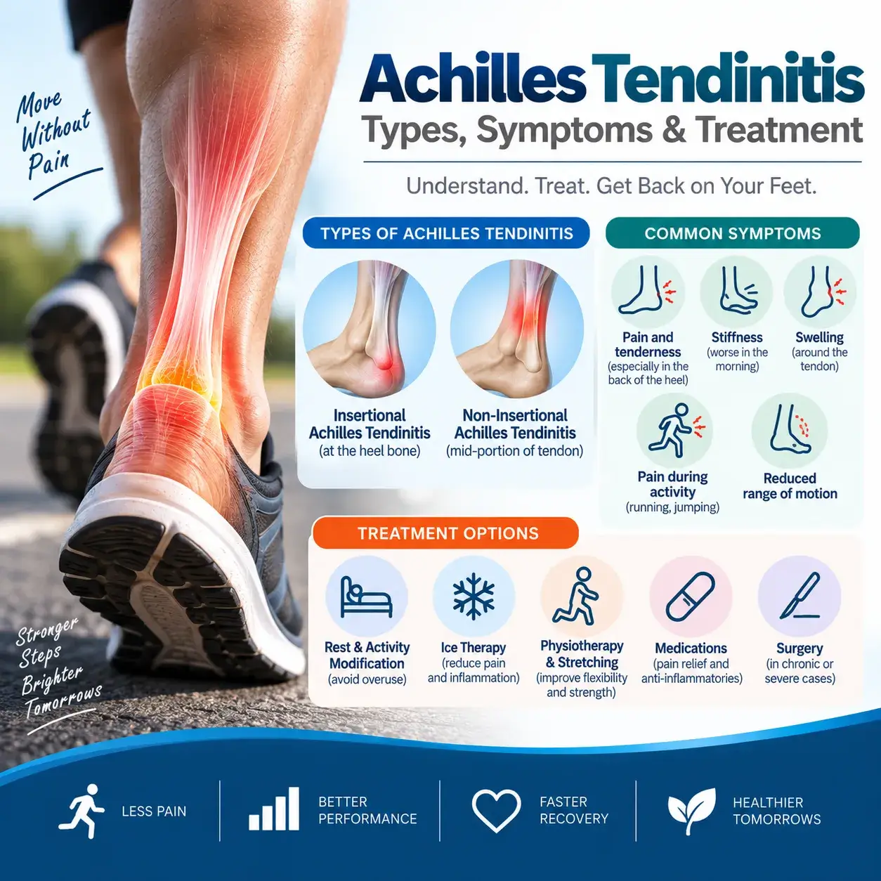
In this type, the fibers in the middle part of the Achilles tendon, just above the point where the tendon attaches to the heel, become affected. Over time, these fibers may deteriorate and develop small tears, which can cause inflammation, swelling, and thickening of the tendon. This type of tendinitis commonly affects younger, physically active individuals, particularly runners.
2. Insertional Achilles Tendinitis
The condition known as Insertional Achilles tendinitis affects the lower part of the tendon where it connects to the heel bone or calcaneus. Over time, the damaged tendon fibers in both insertional and nonintentional Achilles tendinitis can harden (calcify) and form bone spurs on the heel, specifically in the case of insertional Achilles tendinitis. This condition can occur regardless of the activity level or time, although it is more prevalent among runners. Calf muscle tightness is a common cause of this condition as it increases the stress on the Achilles tendon insertion.
When to see a doctor?
If you experience any of the signs and symptoms associated with Achilles tendinitis – and also, experience severe and persistent pain along the Achilles tendon or the back of the heel, then you should see an orthopedic doctor immediately.
Orthopedic Doctor’s Examination
When you see a doctor and describe your symptoms, the doctor will examine your foot and ankle. During the physical examination, your doctor will see whether you feel pain while stretching your calf. The doctor will also look for these signs – swelling at the back of your heel; pain or bone spurs near the lower part pf the tendon; a reduced or restricted ability to point toes downward and pain in the middle of the tendon.
Treatment
Generally, when it comes to Achilles tendinitis, non-surgical treatments can effectively relieve pain, but it may take a few months for symptoms to disappear completely. Even with early treatment, the pain may persist for more than 3 months.
Anti-inflammatory pain medicines, corticosteroid injections, modifying activities, changing footwear, and doing physical therapy exercises are the primary methods used for non-surgical treatment.
Bottom Line
Most instances of Achilles tendinitis can be addressed through basic self-care methods with the oversight of a medical professional. It’s crucial to adhere to these approaches to avoid the condition from happening again. Nevertheless, for severe cases, Achilles tendinitis can lead to torn or ruptured tendons that may necessitate surgical treatment.
Hip Dysplasia Causes &Treatment
Hip Dysplasia Causes
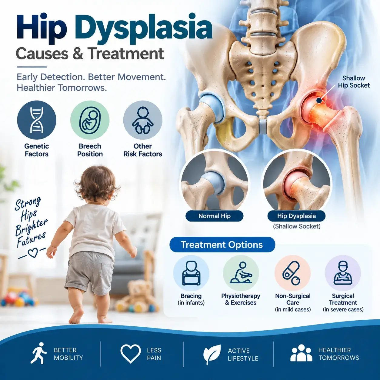
The ball portion of the upper thigh bone fits into the hip socket and forms a stable ball and socket joint for smooth movement. If the socket doesn’t fully cover the ball portion, then the condition is known as hip dysplasia. It damages the cartilage lining the hip joint and the labrum (the soft cartilage) that covers the hip joint’s socket – hip labral tear.
This condition is mostly present in a child shortly after birth. However, if the condition is mild, then it doesn’t cause any symptoms until the child grows into a teenager or young adult.
Hip Dysplasia Symptoms
The signs and symptoms may vary according to the age group. During infancy, the mother can easily notice less flexibility in one hip than the other. The child limps as he or she begins to walk. One leg of the child seems to be longer than the other.
In teenagers and young adults, hip dysplasia can cause painful conditions such as hip labral tear or osteoarthritis. The pain in the hip and groin region increases with activity. Some persons may experience a sensation of instability in the hip joint.
Hip Dysplasia Causes
The problem mostly begins at birth. At this time of a person’s life, the hip joint is made of soft cartilage – which becomes hardened into bone. The ball and socket joint fit together perfectly as a mold. But when the ball fails to fit firmly into the socket – it remains shallow and does not fully form around the ball.
The hip joint moves out from its position during the last month before birth due to the space within the womb becoming crowded. The reduction in space in the womb may be due to the large size of the baby, first pregnancy, and breech presentation.
Hip Dysplasia Risk Factors
The risk increases if babies are born in the breech position. The condition is common in girls and also runs in families.
Complications
Hip dysplasia causes this condition: Hip dysplasia can lead to hip labral tear over time as it damages the labrum (the soft cartilage that rims the hip socket). The risk of osteoarthritis increases in people who have hip dysplasia.
Treatment
For children a variety of treatment options are available. The treatment depends on the extent of hip damage and the age of the child. For mild to moderate cases, orthopedic doctors treat the child with a soft brace. For babies older than 6 months, braces won’t work well. The orthopedic doctor repositions the bone in the proper position and then uses a full body cast to hold the hip joint in a proper position.
However, in severe cases, doctors correct the hip socket by cutting it free from the pelvis and then repositioning it so that it matches perfectly with the ball. The procedure is known as periacetabular osteotomy.
In older people, dysplasia can severely damage their hip joints leading to debilitating arthritis. For older people, hip replacement surgery remains an option.
Bottom Line
In older children, young adults, and older people, orthopedic doctors recommend surgery to move the bones into the proper position for proper movement of the joint.
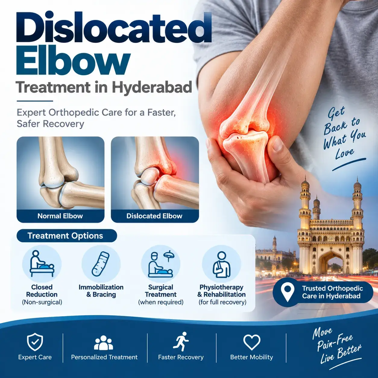
Dislocated elbow treatment in Hyderabad
Elbow Dislocation – Symptoms, Diagnosis & Treatment
Here is the active-voice version, with the passive constructions removed and the grammar cleaned up:
An elbow dislocation occurs when the radius and ulna in the forearm move out of their normal alignment with the humerus in the upper arm. These three bones form the elbow joint, and a dislocation occurs when they lose their normal alignment.
An elbow dislocation can also cause serious injuries, including fractures, damage to the arteries that carry blood to the hand, nerve damage around the elbow, and problems with movement or sensation in the arm and hand.
Causes of Elbow Dislocation
Most elbow dislocations occur after a fall, usually when a person lands with the arm fully extended.
This removes passive constructions such as “are displaced,” “can be dislocated,” “that can occur,” and “are caused by.”
But any trauma, such as a car accident, can cause an elbow dislocation.
Elbow Dislocation Symptoms
- Swelling
- Severe elbow pain
- Weakness in the joint
- Deformed looking arm
- Bruising
- Loss of ability to move the elbow
- Inability to bend the arm
Personal Care at Home
Elbow dislocation is a serious injury that requires medical help. However, you can take some self-care measures at home to ease swelling and pain. For example, you can apply ice to the elbow to help reduce swelling. Afterward, arrange an appointment with an orthopedic doctor for proper evaluation and treatment.
This Simple Test at Home Can Help You Check Whether the Artery and Nerves Are Intact
First, you can check the artery by feeling the base of the wrist below the thumb to locate the pulse. Similarly, press your fingertips gently; they should turn white and return to a pink color within a few seconds. If either test produces an abnormal result, seek medical attention immediately.
Nerve Function Test
In addition, three nerves pass through the elbow, and each nerve plays a role in strength and sensation. First, test your strength by flexing your wrist as if saying “stop” (radial nerve function). Next, extend your fingers to check ulnar nerve function. Finally, try to touch your thumb with your little finger to check median nerve function. If you experience any difficulty during these tests, contact your orthopedic doctor immediately.
Examination and tests
When you visit an orthopedic doctor, the doctor will examine you and perform a physical examination. First, the doctor will check your nerves and arteries by checking your pulse, testing sensation, moving your fingers and wrist, and confirming normal blood flow to your hand.
Next, the doctor will order an X-ray. In some cases, a fracture can look like a dislocation, and a fracture can also occur along with a dislocation.
If your doctor suspects an arterial injury, the doctor may order additional tests, such as an angiogram (X-ray of the arteries).
In addition, the doctor will test sensation by touching different areas of your hand and arm.
Dislocated Elbow Treatment in Hyderabad
During treatment, the doctor will reduce (realign) the elbow by gently retracting the wrist and guiding the joint back into place. Because this procedure can cause significant pain, the doctor may give you strong pain medication before starting the procedure.
When the elbow is back in place, the doctor will take an x-ray and put a splint on it to keep the elbow bent. The splint will form an “L” shape at the back of the elbow. It will be plaster or fiberglass. Its purpose is to prevent the arm from moving through the elbow. Your arms will usually be in a sling to help you hold up your splint.
Prevention
Take precautions and do not walk on slippery stairs and surfaces. Do not fall on outstretched arms. Avoid situations that make falls more common (such as walking at night or on slippery floors). Avoid overuse injuries by restricting overtraining in sports.
When to see a doctor? If you can’t move your elbow, have severe pain, can’t feel your hand, or feel uncomfortable, you should go to your doctor’s office or hospital emergency room right away. For the best dislocated elbow treatment in Hyderabad, meet Dr. Praharsha Mulpur.
